


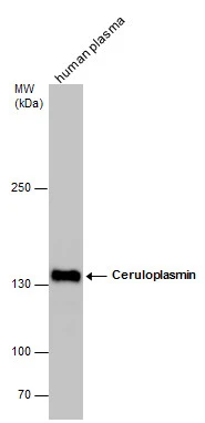
Figure 1. Western blot analysis of Ceruloplasmin using anti-Ceruloplasmin antibody (PB9852). Electrophoresis was performed on a 5-20% SDS-PAGE gel at 70V (Stacking gel) / 90V (Resolving gel) for 2-3 hours. The sample well of each lane was loaded with 30 ug of sample under reducing conditions. Lane 1: 22RV1 whole cell lysates, Lane 2: A549 whole cell lysates. After electrophoresis, proteins were transferred to a nitrocellulose membrane at 150 mA for 50-90 minutes. Blocked the membrane with 5% non-fat milk/TBS for 1.5 hour at RT. The membrane was incubated with rabbit anti-Ceruloplasmin antigen affinity purified polyclonal antibody (Catalog # PB9852) at 0.5 microg/mL overnight at 4°C, then washed with TBS-0.1%Tween 3 times with 5 minutes each and probed with a goat anti-rabbit IgG-HRP secondary antibody at a dilution of 1:5000 for 1.5 hour at RT. The signal is developed using an Enhanced Chemiluminescent detection (ECL) kit (Catalog # EK1002) with Tanon 5200 system. A specific band was detected for Ceruloplasmin at approximately 122 kDa. The expected band size for Ceruloplasmin is at 122 kDa.
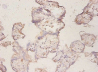
. Ceruloplasmin was detected in a paraffin-embedded section of human liver cancer tissue. Heat mediated antigen retrieval was performed in EDTA buffer (pH 8.0, epitope retrieval solution). The tissue section was blocked with 10% goat serum. The tissue section was then incubated with 1 microg/ml rabbit anti-Ceruloplasmin Antibody (PB9852) overnight at 4°C. Biotinylated goat anti-rabbit IgG was used as secondary antibody and incubated for 30 minutes at 37°C. The tissue section was developed using Strepavidin-Biotin-Complex (SABC) (Catalog # SA1022) with DAB as the chromogen.")
Figure 1. Western blot analysis of Ceruloplasmin using anti-Ceruloplasmin antibody (PB9852). Electrophoresis was performed on a 5-20% SDS-PAGE gel at 70V (Stacking gel) / 90V (Resolving gel) for 2-3 hours. The sample well of each lane was loaded with 30 ug of sample under reducing conditions. Lane 1: 22RV1 whole cell lysates, Lane 2: A549 whole cell lysates. After electrophoresis, proteins were transferred to a nitrocellulose membrane at 150 mA for 50-90 minutes. Blocked the membrane with 5% non-fat milk/TBS for 1.5 hour at RT. The membrane was incubated with rabbit anti-Ceruloplasmin antigen affinity purified polyclonal antibody (Catalog # PB9852) at 0.5 microg/mL overnight at 4°C, then washed with TBS-0.1%Tween 3 times with 5 minutes each and probed with a goat anti-rabbit IgG-HRP secondary antibody at a dilution of 1:5000 for 1.5 hour at RT. The signal is developed using an Enhanced Chemiluminescent detection (ECL) kit (Catalog # EK1002) with Tanon 5200 system. A specific band was detected for Ceruloplasmin at approximately 122 kDa. The expected band size for Ceruloplasmin is at 122 kDa.
Anti-Ceruloplasmin/CP Antibody Picoband(r)
PB9852-CARRIER-FREE
ApplicationsFlow Cytometry, Western Blot
Product group Antibodies
ReactivityHuman
TargetCP
Overview
- SupplierBoster Bio
- Product NameAnti-Ceruloplasmin/CP Antibody Picoband(r)
- Delivery Days Customer9
- Application Supplier NoteTested Species: In-house tested species with positive results. By Heat: Boiling the paraffin sections in 10mM citrate buffer, pH6.0, for 20mins is required for the staining of formalin/paraffin sections. Other applications have not been tested. Optimal dilutions should be determined by end users.
- ApplicationsFlow Cytometry, Western Blot
- CertificationResearch Use Only
- ClonalityPolyclonal
- Concentration500 ug/ml
- Gene ID1356
- Target nameCP
- Target descriptionceruloplasmin
- Target synonymsAB073614, CP-2, ceruloplasmin, caeruloplasmin, ceruloplasmin (ferroxidase), cuproxidase ceruloplasmin, ferroxidase ceruloplasmin, glutathione peroxidase ceruloplasmin, glutathione-dependent peroxiredoxin ceruloplasmin
- HostRabbit
- IsotypeIgG
- Protein IDP00450
- Protein NameCeruloplasmin
- Scientific DescriptionBoster Bio Anti-Ceruloplasmin/CP Antibody Picoband® catalog # PB9852. Tested in Flow Cytometry, WB applications. This antibody reacts with Human. The brand Picoband indicates this is a premium antibody that guarantees superior quality, high affinity, and strong signals with minimal background in Western blot applications. Only our best-performing antibodies are designated as Picoband, ensuring unmatched performance.
- ReactivityHuman
- Storage Instruction-20°C,2°C to 8°C
- UNSPSC12352203
Related products
Product group Antibodies
CP AntibodyCSB-PA14599A0RB
ApplicationsELISA, ImmunoHistoChemistry
ReactivityHuman
TargetCP
- SizePrice
Product group Antibodies
Anti-Ceruloplasmin AntibodyA117071
ApplicationsImmunoPrecipitation
ReactivityHuman
- SizePrice
Product group Antibodies

CP / Ceruloplasmin AntibodyLS-C748698
ApplicationsImmunoHistoChemistry
ReactivityHuman, Mouse, Rat
TargetCP
- SizePrice

Product group Antibodies
Anti-CP AntibodyHPA001834
ApplicationsImmunoHistoChemistry
ReactivityHuman
TargetCP
- SizePrice
Product group Antibodies
Cp Polyclonal AntibodyCAC09179
ApplicationsELISA, ImmunoHistoChemistry
TargetCP
- SizePrice

Product group Antibodies
Ceruloplasmin antibodyGTX101231
ApplicationsWestern Blot, ImmunoHistoChemistry, ImmunoHistoChemistry Paraffin
ReactivityHuman, Mouse, Rat
TargetCP
- SizePrice
Product group Antibodies
Anti-CP Antibody144-07658
ApplicationsWestern Blot
ReactivityHuman, Mouse, Rat
TargetCP
- SizePrice
Product group Antibodies
Ceruloplasmin Recombinant AntibodyBSM-62822R
ApplicationsWestern Blot
ReactivityHuman, Rat
TargetCP
- SizePrice