


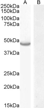
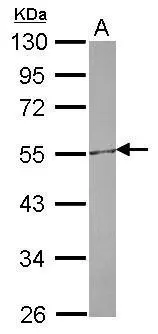
Figure 1. Western blot analysis of CETP using anti-CETP antibody (A00309-1). Electrophoresis was performed on a 5-20% SDS-PAGE gel at 70V (Stacking gel) / 90V (Resolving gel) for 2-3 hours. The sample well of each lane was loaded with 30 ug of sample under reducing conditions. Lane 1: human HCCT tissue lysates, Lane 2: human Hela whole cell lysates, Lane 3: human Jurkat whole cell lysates, Lane 4: rat liver tissue lysates, Lane 5: rat PC-12 whole cell lysates, Lane 6: mouse liver tissue lysates, Lane 7: mouse RAW264.7 whole cell lysates. After electrophoresis, proteins were transferred to a nitrocellulose membrane at 150 mA for 50-90 minutes. Blocked the membrane with 5% non-fat milk/TBS for 1.5 hour at RT. The membrane was incubated with rabbit anti-CETP antigen affinity purified polyclonal antibody (Catalog # A00309-1) at 0.5 microg/mL overnight at 4°C, then washed with TBS-0.1%Tween 3 times with 5 minutes each and probed with a goat anti-rabbit IgG-HRP secondary antibody at a dilution of 1:5000 for 1.5 hour at RT. The signal is developed using an Enhanced Chemiluminescent detection (ECL) kit (Catalog # EK1002) with Tanon 5200 system. A specific band was detected for CETP at approximately 75 kDa. The expected band size for CETP is at 55 kDa.
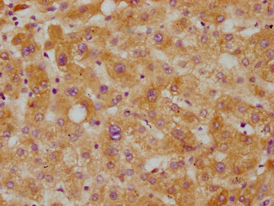
. CETP was detected in paraffin-embedded section of human appendicitis tissues. Heat mediated antigen retrieval was performed in citrate buffer (pH6, epitope retrieval solution) for 20 mins. The tissue section was blocked with 10% goat serum. The tissue section was then incubated with 1microg/ml rabbit anti-CETP Antibody (A00309-1) overnight at 4°C. Biotinylated goat anti-rabbit IgG was used as secondary antibody and incubated for 30 minutes at 37°C. The tissue section was developed using Strepavidin-Biotin-Complex (SABC)(Catalog # SA1022) with DAB as the chromogen.")
. CETP was detected in paraffin-embedded section of human gastric cancer tissues. Heat mediated antigen retrieval was performed in citrate buffer (pH6, epitope retrieval solution) for 20 mins. The tissue section was blocked with 10% goat serum. The tissue section was then incubated with 1microg/ml rabbit anti-CETP Antibody (A00309-1) overnight at 4°C. Biotinylated goat anti-rabbit IgG was used as secondary antibody and incubated for 30 minutes at 37°C. The tissue section was developed using Strepavidin-Biotin-Complex (SABC)(Catalog # SA1022) with DAB as the chromogen.")
. CETP was detected in paraffin-embedded section of human intestinal cancer tissues. Heat mediated antigen retrieval was performed in citrate buffer (pH6, epitope retrieval solution) for 20 mins. The tissue section was blocked with 10% goat serum. The tissue section was then incubated with 1microg/ml rabbit anti-CETP Antibody (A00309-1) overnight at 4°C. Biotinylated goat anti-rabbit IgG was used as secondary antibody and incubated for 30 minutes at 37°C. The tissue section was developed using Strepavidin-Biotin-Complex (SABC)(Catalog # SA1022) with DAB as the chromogen.")
. CETP was detected in paraffin-embedded section of human mammary cancer tissues. Heat mediated antigen retrieval was performed in citrate buffer (pH6, epitope retrieval solution) for 20 mins. The tissue section was blocked with 10% goat serum. The tissue section was then incubated with 1microg/ml rabbit anti-CETP Antibody (A00309-1) overnight at 4°C. Biotinylated goat anti-rabbit IgG was used as secondary antibody and incubated for 30 minutes at 37°C. The tissue section was developed using Strepavidin-Biotin-Complex (SABC)(Catalog # SA1022) with DAB as the chromogen.")
. CETP was detected in paraffin-embedded section of human Ovarian cancer tissues. Heat mediated antigen retrieval was performed in citrate buffer (pH6, epitope retrieval solution) for 20 mins. The tissue section was blocked with 10% goat serum. The tissue section was then incubated with 1microg/ml rabbit anti-CETP Antibody (A00309-1) overnight at 4°C. Biotinylated goat anti-rabbit IgG was used as secondary antibody and incubated for 30 minutes at 37°C. The tissue section was developed using Strepavidin-Biotin-Complex (SABC)(Catalog # SA1022) with DAB as the chromogen.")
. CETP was detected in paraffin-embedded section of human placenta tissues. Heat mediated antigen retrieval was performed in citrate buffer (pH6, epitope retrieval solution) for 20 mins. The tissue section was blocked with 10% goat serum. The tissue section was then incubated with 1microg/ml rabbit anti-CETP Antibody (A00309-1) overnight at 4°C. Biotinylated goat anti-rabbit IgG was used as secondary antibody and incubated for 30 minutes at 37°C. The tissue section was developed using Strepavidin-Biotin-Complex (SABC)(Catalog # SA1022) with DAB as the chromogen.")
. Overlay histogram showing HepG2 cells stained with A00309-1 (Blue line).The cells were blocked with 10% normal goat serum. And then incubated with rabbit anti-CETP Antibody (A00309-1,1microg/1x106 cells) for 30 min at 20°C. DyLight®488 conjugated goat anti-rabbit IgG (BA1127, 5-10microg/1x106 cells) was used as secondary antibody for 30 minutes at 20°C. Isotype control antibody (Green line) was rabbit IgG (1microg/1x106) used under the same conditions. Unlabelled sample (Red line) was also used as a control.")
Figure 1. Western blot analysis of CETP using anti-CETP antibody (A00309-1). Electrophoresis was performed on a 5-20% SDS-PAGE gel at 70V (Stacking gel) / 90V (Resolving gel) for 2-3 hours. The sample well of each lane was loaded with 30 ug of sample under reducing conditions. Lane 1: human HCCT tissue lysates, Lane 2: human Hela whole cell lysates, Lane 3: human Jurkat whole cell lysates, Lane 4: rat liver tissue lysates, Lane 5: rat PC-12 whole cell lysates, Lane 6: mouse liver tissue lysates, Lane 7: mouse RAW264.7 whole cell lysates. After electrophoresis, proteins were transferred to a nitrocellulose membrane at 150 mA for 50-90 minutes. Blocked the membrane with 5% non-fat milk/TBS for 1.5 hour at RT. The membrane was incubated with rabbit anti-CETP antigen affinity purified polyclonal antibody (Catalog # A00309-1) at 0.5 microg/mL overnight at 4°C, then washed with TBS-0.1%Tween 3 times with 5 minutes each and probed with a goat anti-rabbit IgG-HRP secondary antibody at a dilution of 1:5000 for 1.5 hour at RT. The signal is developed using an Enhanced Chemiluminescent detection (ECL) kit (Catalog # EK1002) with Tanon 5200 system. A specific band was detected for CETP at approximately 75 kDa. The expected band size for CETP is at 55 kDa.
Anti-CETP Antibody Picoband(r)
A00309-1-CARRIER-FREE
ApplicationsWestern Blot, ELISA, ImmunoHistoChemistry
Product group Antibodies
ReactivityHuman, Mouse, Rat
TargetCETP
Overview
- SupplierBoster Bio
- Product NameAnti-CETP Antibody Picoband(r)
- Delivery Days Customer9
- ApplicationsWestern Blot, ELISA, ImmunoHistoChemistry
- CertificationResearch Use Only
- ClonalityPolyclonal
- Concentration500 ug/ml
- Gene ID1071
- Target nameCETP
- Target descriptioncholesteryl ester transfer protein
- Target synonymsBPIFF, HDLCQ10, cholesteryl ester transfer protein, BPI fold containing family F, cholesteryl ester transfer protein plasma, lipid transfer protein I
- HostRabbit
- IsotypeIgG
- Protein IDP11597
- Protein NameCholesteryl ester transfer protein
- Scientific DescriptionBoster Bio Anti-CETP Antibody Picoband® catalog # A00309-1. Tested in ELISA, IHC, WB applications. This antibody reacts with Human, Mouse, Rat. The brand Picoband indicates this is a premium antibody that guarantees superior quality, high affinity, and strong signals with minimal background in Western blot applications. Only our best-performing antibodies are designated as Picoband, ensuring unmatched performance.
- ReactivityHuman, Mouse, Rat
- Storage Instruction-20°C,2°C to 8°C
- UNSPSC12352203
Related products
Product group Antibodies
ApplicationsImmunoPrecipitation, Western Blot, ImmunoCytoChemistry, ImmunoHistoChemistry
ReactivityMouse, Porcine
TargetCETP
- SizePrice
Product group Antibodies
Anti-CETP Antibody144-01355
ApplicationsWestern Blot, ImmunoHistoChemistry
ReactivityHuman, Mouse, Rat
TargetCETP
- SizePrice
Product group Antibodies
Anti-CETP AntibodyA286039
ApplicationsWestern Blot, ELISA
ReactivityHuman
- SizePrice
Product group Antibodies

Goat anti-CETPEB09943
ApplicationsWestern Blot, ELISA
ReactivityHuman
TargetCETP
- SizePrice
Product group Antibodies
CETP Recombinant Monoclonal AntibodyCSB-RA005267A0HU
ApplicationsELISA, ImmunoHistoChemistry
ReactivityHuman
TargetCETP
- SizePrice
Product group Antibodies

CETP AntibodyLS-C331424
ApplicationsWestern Blot, ImmunoHistoChemistry
ReactivityHuman
TargetCETP
- SizePrice
Product group Antibodies
CETP Recombinant AntibodyBSM-61156R
ApplicationsImmunoPrecipitation, Western Blot
TargetCETP
- SizePrice
Product group Antibodies
CETP antibody [N1N3]GTX113162
ApplicationsWestern Blot, ImmunoHistoChemistry, ImmunoHistoChemistry Paraffin
ReactivityHuman
TargetCETP
- SizePrice