


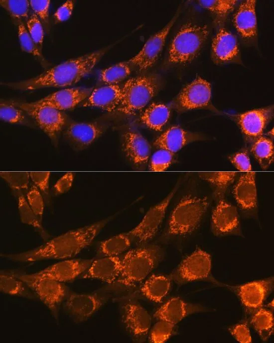
Immunofluorescent staining of human cell line HEL shows localization to vesicles.
Immunofluorescent staining of human cell line HEL shows localization to vesicles.
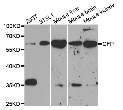
Anti-CFP Antibody
HPA072644
ApplicationsImmunoCytoChemistry
Product group Antibodies
ReactivityHuman
TargetCFP
Overview
- SupplierAtlas Antibodies
- Product NameAnti-CFP Antibody
- Delivery Days Customer4
- ApplicationsImmunoCytoChemistry
- CertificationResearch Use Only
- ClonalityPolyclonal
- ConjugateUnconjugated
- Gene ID5199
- Target nameCFP
- Target descriptioncomplement factor properdin
- Target synonymsBFD, PFC, PFD, PROPERDIN, properdin, complement factor P, properdin P factor, complement
- HostRabbit
- IsotypeIgG
- Protein IDP27918
- Protein NameProperdin
- Scientific DescriptionRecombinant Protein Epitope Signature Tag (PrEST) antigen sequence
- ReactivityHuman
- Storage Instruction-20°C,2°C to 8°C
- UNSPSC41116161
Datasheet
MSDS
Related products
Product group Antibodies
Anti-CFP AntibodyA35670
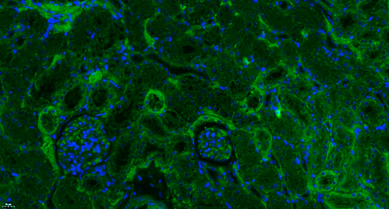
ApplicationsImmunoFluorescence, Western Blot, ImmunoHistoChemistry
ReactivityHuman, Mouse, Rat
- SizePrice
Product group Antibodies

Anti-properdin [3A3E1]AB02512-1.1
ApplicationsWestern Blot, ELISA
ReactivityHuman
TargetCFP
- SizePrice

Product group Antibodies
Anti-CFP Antibody Picoband(r)A00852-2-CARRIER-FREE
ApplicationsFlow Cytometry, Western Blot, ImmunoHistoChemistry, ImmunoHistoChemistry Frozen
ReactivityHuman, Mouse, Rat
TargetCFP
- SizePrice
Product group Antibodies
References
Properdin Polyclonal AntibodyBS-8306R
ApplicationsImmunoFluorescence, Western Blot, ELISA, ImmunoHistoChemistry, ImmunoHistoChemistry Paraffin
ReactivityHuman, Mouse, Rabbit, Rat
TargetCFP
- SizePrice
Product group Antibodies
ApplicationsImmunoPrecipitation, Western Blot, ImmunoCytoChemistry, ImmunoHistoChemistry
TargetCFP
- SizePrice
Product group Antibodies

Goat anti Human properdinGAHu/PPD
ApplicationsImmunoElectrophoresis, ImmunoPrecipitation
TargetCFP
- SizePrice
Product group Antibodies
Properdin antibodyGTX53957
ApplicationsImmunoFluorescence, Western Blot, ImmunoCytoChemistry
ReactivityHuman, Mouse
TargetCFP
- SizePrice
Product group Antibodies
Anti-CFP AntibodyCAB5398
ApplicationsImmunoFluorescence, Western Blot, ELISA, ImmunoCytoChemistry, ImmunoHistoChemistry, ImmunoHistoChemistry Paraffin
ReactivityHuman
TargetCFP
- SizePrice
Product group Antibodies

Properdin / CFP Antibody (aa315-469)LS-C293499
ApplicationsWestern Blot
ReactivityHuman
TargetCFP
- SizePrice