


Immunofluorescent staining of human cell line PC-3 shows localization to mitochondria.
Immunofluorescent staining of human cell line PC-3 shows localization to mitochondria.
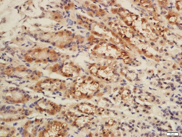
Anti-CISD1 Antibody
HPA074383
ApplicationsImmunoCytoChemistry
Product group Antibodies
ReactivityHuman
TargetCISD1
Overview
- SupplierAtlas Antibodies
- Product NameAnti-CISD1 Antibody
- Delivery Days Customer4
- ApplicationsImmunoCytoChemistry
- CertificationResearch Use Only
- ClonalityPolyclonal
- ConjugateUnconjugated
- Gene ID55847
- Target nameCISD1
- Target descriptionCDGSH iron sulfur domain 1
- Target synonymsC10orf70, MDS029, ZCD1, mitoNEET, CDGSH iron-sulfur domain-containing protein 1, cysteine transaminase CISD1, zinc finger CDGSH-type domain 1
- HostRabbit
- IsotypeIgG
- Protein IDQ9NZ45
- Protein NameCDGSH iron-sulfur domain-containing protein 1
- Scientific DescriptionRecombinant Protein Epitope Signature Tag (PrEST) antigen sequence
- ReactivityHuman
- Storage Instruction-20°C,2°C to 8°C
- UNSPSC41116161
Datasheet
MSDS
Related products

Product group Antibodies
Anti-MitoNEET/CISD1 Antibody Picoband(r)A04360-2-CARRIER-FREE
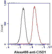
ApplicationsFlow Cytometry, Western Blot, ELISA, ImmunoHistoChemistry
ReactivityHuman, Monkey, Mouse, Rat
TargetCISD1
- SizePrice
Product group Antibodies
Anti-CISD1 Antibody144-10317
ApplicationsWestern Blot
ReactivityHuman, Mouse, Rat
TargetCISD1
- SizePrice
Product group Antibodies

CISD1 AntibodyLS-C668360
ApplicationsWestern Blot
ReactivityHuman
TargetCISD1
- SizePrice
Product group Antibodies
CISD1 AntibodyCSB-PA005442GA01HU
ApplicationsImmunoFluorescence, Western Blot, ELISA, ImmunoHistoChemistry
ReactivityHuman, Mouse, Rat, Zebra Fish
TargetCISD1
- SizePrice
Product group Antibodies
CISD1 Polyclonal AntibodyBS-13959R
ApplicationsImmunoFluorescence, Western Blot, ELISA, ImmunoCytoChemistry, ImmunoHistoChemistry, ImmunoHistoChemistry Frozen, ImmunoHistoChemistry Paraffin
ReactivityBovine, Canine, Equine, Human, Mouse, Rabbit, Rat, Sheep
TargetCISD1
- SizePrice
Product group Antibodies
CISD1 antibody [AT1A8]GTX57586
ApplicationsFlow Cytometry, Western Blot
ReactivityHuman, Mouse
TargetCISD1
- SizePrice