

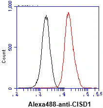
FACS analysis of THP-1 cells using GTX57586 CISD1 antibody. Cell Number: 1 x 10? cells Primary antibody: Red line Antibody amount: 2-5 μg


FACS analysis of THP-1 cells using GTX57586 CISD1 antibody. Cell Number: 1 x 10? cells Primary antibody: Red line Antibody amount: 2-5 μg
CISD1 antibody [AT1A8]
GTX57586
ApplicationsFlow Cytometry, Western Blot
Product group Antibodies
ReactivityHuman, Mouse
TargetCISD1
Overview
- SupplierGeneTex
- Product NameCISD1 antibody [AT1A8]
- Delivery Days Customer9
- Application Supplier NoteWB: Recommended starting dilution is 1:1000.. *Optimal dilutions/concentrations should be determined by the researcher.Not tested in other applications.
- ApplicationsFlow Cytometry, Western Blot
- CertificationResearch Use Only
- ClonalityMonoclonal
- Clone IDAT1A8
- Concentration1 mg/ml
- ConjugateUnconjugated
- Gene ID55847
- Target nameCISD1
- Target descriptionCDGSH iron sulfur domain 1
- Target synonymsC10orf70, MDS029, ZCD1, mitoNEET, CDGSH iron-sulfur domain-containing protein 1, cysteine transaminase CISD1, zinc finger CDGSH-type domain 1
- HostMouse
- IsotypeIgG1
- Protein IDQ9NZ45
- Protein NameCDGSH iron-sulfur domain-containing protein 1
- Scientific DescriptionThis gene encodes a protein with a CDGSH iron-sulfur domain and has been shown to bind a redox-active [2Fe-2S] cluster. The encoded protein has been localized to the outer membrane of mitochondria and is thought to play a role in regulation of oxidation. Genes encoding similar proteins are located on chromosomes 4 and 17, and a pseudogene of this gene is located on chromosome 2. [provided by RefSeq, Feb 2012]
- ReactivityHuman, Mouse
- Storage Instruction-20°C or -80°C,2°C to 8°C
- UNSPSC41116161
Datasheet
Related products

Product group Antibodies
Anti-MitoNEET/CISD1 Antibody Picoband(r)A04360-2-CARRIER-FREE
ApplicationsFlow Cytometry, Western Blot, ELISA, ImmunoHistoChemistry
ReactivityHuman, Monkey, Mouse, Rat
TargetCISD1
- SizePrice
Product group Antibodies
Anti-CISD1 Antibody144-10317
ApplicationsWestern Blot
ReactivityHuman, Mouse, Rat
TargetCISD1
- SizePrice
Product group Antibodies

CISD1 AntibodyLS-C668360
ApplicationsWestern Blot
ReactivityHuman
TargetCISD1
- SizePrice
Product group Antibodies
CISD1 AntibodyCSB-PA005442GA01HU
ApplicationsImmunoFluorescence, Western Blot, ELISA, ImmunoHistoChemistry
ReactivityHuman, Mouse, Rat, Zebra Fish
TargetCISD1
- SizePrice
Product group Antibodies
CISD1 Polyclonal AntibodyBS-13959R
ApplicationsImmunoFluorescence, Western Blot, ELISA, ImmunoCytoChemistry, ImmunoHistoChemistry, ImmunoHistoChemistry Frozen, ImmunoHistoChemistry Paraffin
ReactivityBovine, Canine, Equine, Human, Mouse, Rabbit, Rat, Sheep
TargetCISD1
- SizePrice
![FACS analysis of Jurkat cells using GTX84683 CISD1 antibody [2B3]. Red : Primary antibody Blue : Negative control antibody](https://www.genetex.com/upload/website/prouct_img/normal/GTX84683/GTX84683_503_FACS_w_23061420_137.webp)
Product group Antibodies
CISD1 antibody [2B3]GTX84683
ApplicationsFlow Cytometry, ImmunoFluorescence, Western Blot, ImmunoCytoChemistry
ReactivityCanine, Human
TargetCISD1
- SizePrice

Product group Antibodies
Anti-CISD1 AntibodyHPA074383
ApplicationsImmunoCytoChemistry
ReactivityHuman
TargetCISD1
- SizePrice
Product group Antibodies
CISD1 antibodyGTX65611
ApplicationsWestern Blot
ReactivityHuman, Mouse, Rat
TargetCISD1
- SizePrice