


Immunohistochemical staining of human cerebral cortex shows strong cytoplasmic positivity in neurons.
Immunohistochemical staining of human cerebral cortex shows strong cytoplasmic positivity in neurons.
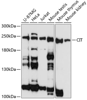
Anti-CIT Antibody
HPA019082
ApplicationsImmunoHistoChemistry
Product group Antibodies
ReactivityHuman
TargetCIT
Overview
- SupplierAtlas Antibodies
- Product NameAnti-CIT Antibody
- Delivery Days Customer4
- ApplicationsImmunoHistoChemistry
- CertificationResearch Use Only
- ClonalityPolyclonal
- ConjugateUnconjugated
- Gene ID11113
- Target nameCIT
- Target descriptioncitron rho-interacting serine/threonine kinase
- Target synonymsCITK, CRIK, MCPH17, STK21, citron Rho-interacting kinase, citron (rho-interacting, serine/threonine kinase 21), serine/threonine kinase 21, serine/threonine-protein kinase 21
- HostRabbit
- IsotypeIgG
- Protein IDO14578
- Protein NameCitron Rho-interacting kinase
- Scientific DescriptionRecombinant Protein Epitope Signature Tag (PrEST) antigen sequence
- ReactivityHuman
- Storage Instruction-20°C,2°C to 8°C
- UNSPSC41116161
Datasheet
MSDS
Related products
Product group Antibodies
Anti-CIT AntibodyA88805
ApplicationsWestern Blot
ReactivityHuman, Mouse
- SizePrice

Product group Antibodies
Anti-CIT Antibody Picoband(r)A01331-2-CARRIER-FREE
ApplicationsFlow Cytometry, Western Blot, ELISA
ReactivityHuman, Mouse, Rat
TargetCIT
- SizePrice
Product group Antibodies
CIT AntibodyCSB-PA006777
ApplicationsELISA, ImmunoHistoChemistry
ReactivityHuman, Mouse
TargetCIT
- SizePrice
Product group Antibodies

ApplicationsImmunoPrecipitation, Western Blot
ReactivityHuman
TargetCIT
- SizePrice

Product group Antibodies
Anti-CIT AntibodyHPA048812
ApplicationsImmunoHistoChemistry
ReactivityHuman
TargetCIT
- SizePrice

Product group Antibodies
Anti-CIT AntibodyHPA075506
ApplicationsImmunoCytoChemistry
ReactivityHuman
TargetCIT
- SizePrice