


Figure 1. Flow Cytometry analysis of Jurkat cells using anti-CLEC9A antibody (A04577). Overlay histogram showing Jurkat cells stained with A04577 (Blue line). To facilitate intracellular staining, cells were fixed with 4% paraformaldehyde and permeabilized with permeabilization buffer. The cells were blocked with 10% normal goat serum. And then incubated with rabbit anti-CLEC9A Antibody (A04577, 1microg/1x106 cells) for 30 min at 20°C. DyLight®488 conjugated goat anti-rabbit IgG (BA1127, 5-10microg/1x106 cells) was used as secondary antibody for 30 minutes at 20°C. Isotype control antibody (Green line) was rabbit IgG (1microg/1x106) used under the same conditions. Unlabelled sample (Red line) was also used as a control.
Figure 1. Flow Cytometry analysis of Jurkat cells using anti-CLEC9A antibody (A04577). Overlay histogram showing Jurkat cells stained with A04577 (Blue line). To facilitate intracellular staining, cells were fixed with 4% paraformaldehyde and permeabilized with permeabilization buffer. The cells were blocked with 10% normal goat serum. And then incubated with rabbit anti-CLEC9A Antibody (A04577, 1microg/1x106 cells) for 30 min at 20°C. DyLight®488 conjugated goat anti-rabbit IgG (BA1127, 5-10microg/1x106 cells) was used as secondary antibody for 30 minutes at 20°C. Isotype control antibody (Green line) was rabbit IgG (1microg/1x106) used under the same conditions. Unlabelled sample (Red line) was also used as a control.
Anti-CLEC9A Antibody
A04577-CARRIER-FREE
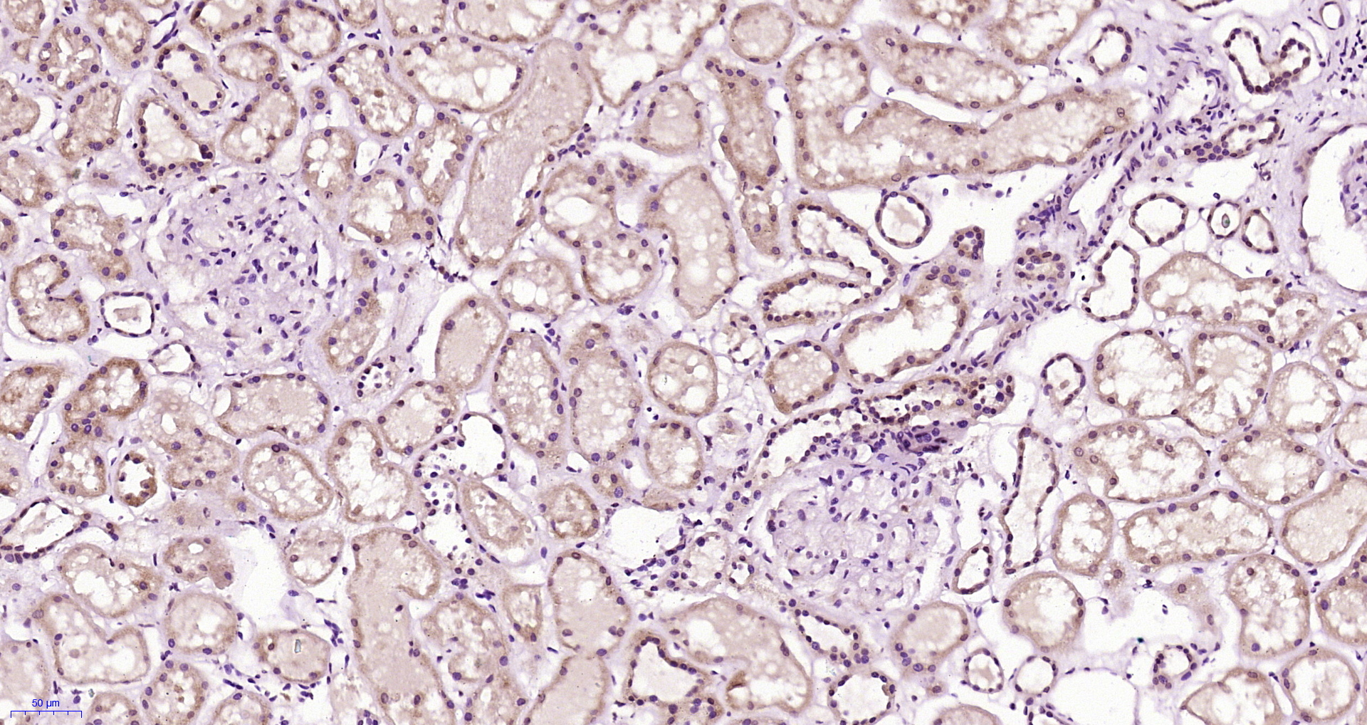
ApplicationsFlow Cytometry, ImmunoCytoChemistry, ImmunoHistoChemistry
Product group Antibodies
ReactivityHuman
TargetCLEC9A
Overview
- SupplierBoster Bio
- Product NameAnti-CLEC9A Antibody
- Delivery Days Customer9
- ApplicationsFlow Cytometry, ImmunoCytoChemistry, ImmunoHistoChemistry
- CertificationResearch Use Only
- ClonalityPolyclonal
- Concentration500 ug/ml
- Gene ID283420
- Target nameCLEC9A
- Target descriptionC-type lectin domain containing 9A
- Target synonymsCD370, DNGR-1, DNGR1, UNQ9341, C-type lectin domain family 9 member A, HEEE9341
- HostRabbit
- IsotypeIgG
- Protein IDQ6UXN8
- Protein NameC-type lectin domain family 9 member A
- Scientific DescriptionBoster Bio Anti-CLEC9A Antibody Picoband® catalog # A04577. Tested in Flow Cytometry, IHC, ICC applications. This antibody reacts with Human.
- ReactivityHuman
- Storage Instruction-20°C,2°C to 8°C
- UNSPSC12352203
Related products
Product group Antibodies

CLEC9A AntibodyLS-C832660
ApplicationsWestern Blot, ELISA
ReactivityHuman
TargetCLEC9A
- SizePrice
Product group Antibodies
CLEC9A Polyclonal AntibodyBS-13620R
ApplicationsImmunoFluorescence, Western Blot, ELISA, ImmunoCytoChemistry, ImmunoHistoChemistry, ImmunoHistoChemistry Frozen, ImmunoHistoChemistry Paraffin
ReactivityHuman, Mouse, Rat
TargetCLEC9A
- SizePrice
Product group Antibodies
CLEC9A Recombinant Monoclonal AntibodyCSB-RA740922MA1HU
ApplicationsELISA
ReactivityHuman
TargetCLEC9A
- SizePrice
Product group Antibodies
CLEC9A Polyclonal AntibodyCAC15930
ApplicationsImmunoFluorescence, Western Blot, ELISA
ReactivityMouse
TargetCLEC9A
- SizePrice
Product group Antibodies
CLEC9A antibody [30L2]GTX53076
ApplicationsFlow Cytometry
ReactivityHuman
TargetCLEC9A
- SizePrice