


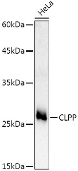
Figure 1. Western blot analysis of CLPP using anti-CLPP antibody (A01117-1). Electrophoresis was performed on a 5-20% SDS-PAGE gel at 70V (Stacking gel) / 90V (Resolving gel) for 2-3 hours. The sample well of each lane was loaded with 30ug of sample under reducing conditions. Lane 1: human PC-3 whole cell lysates, Lane 2: human HEK293 whole cell lysates, Lane 3: human MCF-7 whole cell lysates, Lane 4: human THP-1 whole cell lysates, Lane 5: rat pancreas tissue lysates, Lane 6: rat lung tissue lysates, Lane 7: mouse pancreas tissue lysates. After Electrophoresis, proteins were transferred to a Nitrocellulose membrane at 150mA for 50-90 minutes. Blocked the membrane with 5% Non-fat Milk/ TBS for 1.5 hour at RT. The membrane was incubated with rabbit anti-CLPP antigen affinity purified polyclonal antibody (Catalog # A01117-1) at 0.5 microg/mL overnight at 4°C, then washed with TBS-0.1%Tween 3 times with 5 minutes each and probed with a goat anti-rabbit IgG-HRP secondary antibody at a dilution of 1:5000 for 1.5 hour at RT. The signal is developed using an Enhanced Chemiluminescent detection (ECL) kit (Catalog # EK1002) with Tanon 5200 system. A specific band was detected for CLPP at approximately 26KD. The expected band size for CLPP is at 26KD.
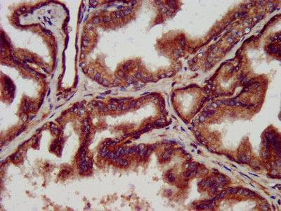
. CLPP was detected in paraffin-embedded section of human breast cancer tissue. Heat mediated antigen retrieval was performed in EDTA buffer (pH8.0, epitope retrieval solution). The tissue section was blocked with 10% goat serum. The tissue section was then incubated with 1microg/ml rabbit anti-CLPP Antibody (A01117-1) overnight at 4°C. Biotinylated goat anti-rabbit IgG was used as secondary antibody and incubated for 30 minutes at 37°C. The tissue section was developed using Strepavidin-Biotin-Complex (SABC) (Catalog # SA1022) with DAB as the chromogen.")
. CLPP was detected in paraffin-embedded section of human liver cancer tissue. Heat mediated antigen retrieval was performed in EDTA buffer (pH8.0, epitope retrieval solution). The tissue section was blocked with 10% goat serum. The tissue section was then incubated with 1microg/ml rabbit anti-CLPP Antibody (A01117-1) overnight at 4°C. Biotinylated goat anti-rabbit IgG was used as secondary antibody and incubated for 30 minutes at 37°C. The tissue section was developed using Strepavidin-Biotin-Complex (SABC) (Catalog # SA1022) with DAB as the chromogen.")
. CLPP was detected in paraffin-embedded section of human lung cancer tissue. Heat mediated antigen retrieval was performed in EDTA buffer (pH8.0, epitope retrieval solution). The tissue section was blocked with 10% goat serum. The tissue section was then incubated with 1microg/ml rabbit anti-CLPP Antibody (A01117-1) overnight at 4°C. Biotinylated goat anti-rabbit IgG was used as secondary antibody and incubated for 30 minutes at 37°C. The tissue section was developed using Strepavidin-Biotin-Complex (SABC) (Catalog # SA1022) with DAB as the chromogen.")
. CLPP was detected in paraffin-embedded section of human ovarian cancer tissue. Heat mediated antigen retrieval was performed in EDTA buffer (pH8.0, epitope retrieval solution). The tissue section was blocked with 10% goat serum. The tissue section was then incubated with 1microg/ml rabbit anti-CLPP Antibody (A01117-1) overnight at 4°C. Biotinylated goat anti-rabbit IgG was used as secondary antibody and incubated for 30 minutes at 37°C. The tissue section was developed using Strepavidin-Biotin-Complex (SABC) (Catalog # SA1022) with DAB as the chromogen.")
. CLPP was detected in paraffin-embedded section of human gallbladder adenocarcinoma tissue. Heat mediated antigen retrieval was performed in EDTA buffer (pH8.0, epitope retrieval solution). The tissue section was blocked with 10% goat serum. The tissue section was then incubated with 1microg/ml rabbit anti-CLPP Antibody (A01117-1) overnight at 4°C. Biotinylated goat anti-rabbit IgG was used as secondary antibody and incubated for 30 minutes at 37°C. The tissue section was developed using Strepavidin-Biotin-Complex (SABC) (Catalog # SA1022) with DAB as the chromogen.")
. CLPP was detected in immunocytochemical section of MCF-7 cells. Enzyme antigen retrieval was performed using IHC enzyme antigen retrieval reagent (AR0022) for 15 mins. The cells were blocked with 10% goat serum. And then incubated with 2microg/mL rabbit anti-CLPP Antibody (A01117-1) overnight at 4°C. DyLight®488 Conjugated Goat Anti-Rabbit IgG (BA1127) was used as secondary antibody at 1:100 dilution and incubated for 30 minutes at 37°C. The section was counterstained with DAPI. Visualize using a fluorescence microscope and filter sets appropriate for the label used.")
. Overlay histogram showing U937 cells stained with A01117-1 (Blue line). To facilitate intracellular staining, cells were fixed with 4% paraformaldehyde and permeabilized with permeabilization buffer. The cells were blocked with 10% normal goat serum. And then incubated with rabbit anti-CLPP Antibody (A01117-1, 1microg/1x106 cells) for 30 min at 20°C. DyLight®488 conjugated goat anti-rabbit IgG (BA1127, 5-10microg/1x106 cells) was used as secondary antibody for 30 minutes at 20°C. Isotype control antibody (Green line) was rabbit IgG (1microg/1x106) used under the same conditions. Unlabelled sample without incubation with primary antibody and secondary antibody (Red line) was used as a blank control.")
Figure 1. Western blot analysis of CLPP using anti-CLPP antibody (A01117-1). Electrophoresis was performed on a 5-20% SDS-PAGE gel at 70V (Stacking gel) / 90V (Resolving gel) for 2-3 hours. The sample well of each lane was loaded with 30ug of sample under reducing conditions. Lane 1: human PC-3 whole cell lysates, Lane 2: human HEK293 whole cell lysates, Lane 3: human MCF-7 whole cell lysates, Lane 4: human THP-1 whole cell lysates, Lane 5: rat pancreas tissue lysates, Lane 6: rat lung tissue lysates, Lane 7: mouse pancreas tissue lysates. After Electrophoresis, proteins were transferred to a Nitrocellulose membrane at 150mA for 50-90 minutes. Blocked the membrane with 5% Non-fat Milk/ TBS for 1.5 hour at RT. The membrane was incubated with rabbit anti-CLPP antigen affinity purified polyclonal antibody (Catalog # A01117-1) at 0.5 microg/mL overnight at 4°C, then washed with TBS-0.1%Tween 3 times with 5 minutes each and probed with a goat anti-rabbit IgG-HRP secondary antibody at a dilution of 1:5000 for 1.5 hour at RT. The signal is developed using an Enhanced Chemiluminescent detection (ECL) kit (Catalog # EK1002) with Tanon 5200 system. A specific band was detected for CLPP at approximately 26KD. The expected band size for CLPP is at 26KD.
Anti-CLPP Antibody Picoband(r)
A01117-1-CARRIER-FREE
ApplicationsFlow Cytometry, ImmunoFluorescence, Western Blot, ELISA, ImmunoCytoChemistry, ImmunoHistoChemistry
Product group Antibodies
ReactivityHuman, Mouse, Rat
TargetCLPP
Overview
- SupplierBoster Bio
- Product NameAnti-CLPP Antibody Picoband(r)
- Delivery Days Customer9
- ApplicationsFlow Cytometry, ImmunoFluorescence, Western Blot, ELISA, ImmunoCytoChemistry, ImmunoHistoChemistry
- CertificationResearch Use Only
- ClonalityPolyclonal
- Concentration500 ug/ml
- Gene ID8192
- Target nameCLPP
- Target descriptioncaseinolytic mitochondrial matrix peptidase proteolytic subunit
- Target synonymsDFNB81, PRLTS3, ATP-dependent Clp protease proteolytic subunit, mitochondrial, ATP-dependent protease ClpAP, proteolytic subunit, human, ClpP caseinolytic peptidase ATP-dependent, proteolytic subunit, ClpP caseinolytic peptidase, ATP-dependent, proteolytic subunit homolog, ClpP caseinolytic protease, ATP-dependent, proteolytic subunit homolog, endopeptidase Clp, putative ATP-dependent Clp protease proteolytic subunit, mitochondrial
- HostRabbit
- IsotypeIgG
- Protein IDQ16740
- Protein NameATP-dependent Clp protease proteolytic subunit, mitochondrial
- Scientific DescriptionBoster Bio Anti-CLPP Antibody Picoband® catalog # A01117-1. Tested in ELISA, Flow Cytometry, IF, IHC, ICC, WB applications. This antibody reacts with Human, Mouse, Rat. The brand Picoband indicates this is a premium antibody that guarantees superior quality, high affinity, and strong signals with minimal background in Western blot applications. Only our best-performing antibodies are designated as Picoband, ensuring unmatched performance.
- ReactivityHuman, Mouse, Rat
- Storage Instruction-20°C,2°C to 8°C
- UNSPSC12352203
Related products
Product group Antibodies
CLPP AntibodyCSB-PA618094LA01HU
ApplicationsELISA, ImmunoHistoChemistry
ReactivityHuman
TargetCLPP
- SizePrice
Product group Antibodies
Anti-CLPP AntibodyA16297
ApplicationsWestern Blot
ReactivityHuman, Mouse, Rat
- SizePrice
Product group Antibodies

Goat anti-CLPPEB05873
ApplicationsWestern Blot, ELISA, ImmunoHistoChemistry
ReactivityHuman
TargetCLPP
- SizePrice

Product group Antibodies
Anti-CLPP AntibodyHPA010649
ApplicationsWestern Blot, ImmunoCytoChemistry, ImmunoHistoChemistry
ReactivityHuman, Mouse, Rat
TargetCLPP
- SizePrice
Product group Antibodies

CLPP AntibodyLS-C410661
ApplicationsWestern Blot
ReactivityHuman, Mouse, Rat
TargetCLPP
- SizePrice
Product group Antibodies
CLPP Polyclonal AntibodyCAC13345
ApplicationsELISA, ImmunoHistoChemistry
TargetCLPP
- SizePrice
![CLPP antibody [C2C3], C-term detects CLPP protein at mitochondria by immunofluorescent analysis. Sample: A431 cells were fixed in 2% paraformaldehyde/culture medium at RT for 30 min. Green: CLPP protein stained by CLPP antibody [C2C3], C-term (GTX104656) diluted at 1:500. Blue: Hoechst 33342 staining. Scale bar = 10 μm.](https://www.genetex.com/upload/website/prouct_img/normal/GTX104656/GTX104656_41752_20150629_IFA_w_23060120_763.webp)
Product group Antibodies
CLPP antibody [C2C3], C-termGTX104656
ApplicationsImmunoFluorescence, Western Blot, ImmunoCytoChemistry, ImmunoHistoChemistry, ImmunoHistoChemistry Paraffin
ReactivityHuman
TargetCLPP
- SizePrice
Product group Antibodies
ApplicationsFlow Cytometry, Western Blot, ImmunoCytoChemistry
ReactivityHuman, Mouse, Rat
TargetCLPP
- SizePrice
Product group Antibodies
Anti-CLPP Antibody144-09127
ApplicationsWestern Blot
ReactivityHuman, Mouse, Rat
TargetCLPP
- SizePrice