


Figure 1. Western blot analysis of PTGS2 using anti-PTGS2 antibody (A00084-2). Electrophoresis was performed on a 5-20% SDS-PAGE gel at 70V (Stacking gel) / 90V (Resolving gel) for 2-3 hours. The sample well of each lane was loaded with 30 ug of sample under reducing conditions. Lane 1: human Hela whole cell lysates, Lane 2: mouse RAW264.7(-LPS) whole cell lysates, Lane 3: mouse RAW264.7(+LPS) whole cell lysates. After electrophoresis, proteins were transferred to a nitrocellulose membrane at 150 mA for 50-90 minutes. Blocked the membrane with 5% non-fat milk/TBS for 1.5 hour at RT. The membrane was incubated with rabbit anti-PTGS2 antigen affinity purified polyclonal antibody (Catalog # A00084-2) at 0.25 microg/mL overnight at 4°C, then washed with TBS-0.1%Tween 3 times with 5 minutes each and probed with a goat anti-rabbit IgG-HRP secondary antibody at a dilution of 1:5000 for 1.5 hour at RT. The signal is developed using an Enhanced Chemiluminescent detection (ECL) kit (Catalog # EK1002) with Tanon 5200 system. A specific band was detected for PTGS2 at approximately 75 kDa. The expected band size for PTGS2 is at 69 kDa.
. PTGS2 was detected in a paraffin-embedded section of human lung cancer tissue. Heat mediated antigen retrieval was performed in EDTA buffer (pH 8.0, epitope retrieval solution). The tissue section was blocked with 10% goat serum. The tissue section was then incubated with 2 microg/ml rabbit anti-PTGS2 Antibody (A00084-2) overnight at 4°C. Peroxidase Conjugated Goat Anti-rabbit IgG was used as secondary antibody and incubated for 30 minutes at 37°C. The tissue section was developed using HRP Conjugated Rabbit IgG Super Vision Assay Kit (Catalog # SV0002) with DAB as the chromogen.")
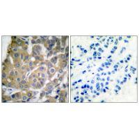
. PTGS2 was detected in a paraffin-embedded section of human pancreatic cancer tissue. Heat mediated antigen retrieval was performed in EDTA buffer (pH 8.0, epitope retrieval solution). The tissue section was blocked with 10% goat serum. The tissue section was then incubated with 2 microg/ml rabbit anti-PTGS2 Antibody (A00084-2) overnight at 4°C. Peroxidase Conjugated Goat Anti-rabbit IgG was used as secondary antibody and incubated for 30 minutes at 37°C. The tissue section was developed using HRP Conjugated Rabbit IgG Super Vision Assay Kit (Catalog # SV0002) with DAB as the chromogen.")
. Overlay histogram showing CACO-2 cells stained with A00084-2 (Blue line). To facilitate intracellular staining, cells were fixed with 4% paraformaldehyde and permeabilized with permeabilization buffer. The cells were blocked with 10% normal goat serum. And then incubated with rabbit anti-PTGS2 Antibody (A00084-2, 1microg/1x106 cells) for 30 min at 20°C. DyLight®488 conjugated goat anti-rabbit IgG (BA1127, 5-10microg/1x106 cells) was used as secondary antibody for 30 minutes at 20°C. Isotype control antibody (Green line) was rabbit IgG (1microg/1x106) used under the same conditions. Unlabelled sample (Red line) was also used as a control.")
. Overlay histogram showing HEPA1-6 cells stained with A00084-2 (Blue line). To facilitate intracellular staining, cells were fixed with 4% paraformaldehyde and permeabilized with permeabilization buffer. The cells were blocked with 10% normal goat serum. And then incubated with rabbit anti-PTGS2 Antibody (A00084-2, 1microg/1x106 cells) for 30 min at 20°C. DyLight®488 conjugated goat anti-rabbit IgG (BA1127, 5-10microg/1x106 cells) was used as secondary antibody for 30 minutes at 20°C. Isotype control antibody (Green line) was rabbit IgG (1microg/1x106) used under the same conditions. Unlabelled sample (Red line) was also used as a control.")
Figure 1. Western blot analysis of PTGS2 using anti-PTGS2 antibody (A00084-2). Electrophoresis was performed on a 5-20% SDS-PAGE gel at 70V (Stacking gel) / 90V (Resolving gel) for 2-3 hours. The sample well of each lane was loaded with 30 ug of sample under reducing conditions. Lane 1: human Hela whole cell lysates, Lane 2: mouse RAW264.7(-LPS) whole cell lysates, Lane 3: mouse RAW264.7(+LPS) whole cell lysates. After electrophoresis, proteins were transferred to a nitrocellulose membrane at 150 mA for 50-90 minutes. Blocked the membrane with 5% non-fat milk/TBS for 1.5 hour at RT. The membrane was incubated with rabbit anti-PTGS2 antigen affinity purified polyclonal antibody (Catalog # A00084-2) at 0.25 microg/mL overnight at 4°C, then washed with TBS-0.1%Tween 3 times with 5 minutes each and probed with a goat anti-rabbit IgG-HRP secondary antibody at a dilution of 1:5000 for 1.5 hour at RT. The signal is developed using an Enhanced Chemiluminescent detection (ECL) kit (Catalog # EK1002) with Tanon 5200 system. A specific band was detected for PTGS2 at approximately 75 kDa. The expected band size for PTGS2 is at 69 kDa.
Anti-COX2/Cyclooxygenase 2/PTGS2 Picoband(r) Antibody
A00084-2-CARRIER-FREE
ApplicationsFlow Cytometry, Western Blot, ELISA, ImmunoHistoChemistry
Product group Antibodies
ReactivityHuman, Mouse
TargetPTGS2
Overview
- SupplierBoster Bio
- Product NameAnti-COX2/Cyclooxygenase 2/PTGS2 Picoband(r) Antibody
- Delivery Days Customer9
- Application Supplier NoteTested Species: In-house tested species with positive results. Other applications have not been tested. Optimal dilutions should be determined by end users.
- ApplicationsFlow Cytometry, Western Blot, ELISA, ImmunoHistoChemistry
- CertificationResearch Use Only
- ClonalityPolyclonal
- Concentration500 ug/ml
- Gene ID5743
- Target namePTGS2
- Target descriptionprostaglandin-endoperoxide synthase 2
- Target synonymsCOX-2, COX2, GRIPGHS, PGG/HS, PGHS-2, PHS-2, hCox-2, prostaglandin G/H synthase 2, PGH synthase 2, PHS II, cyclooxygenase 2, cyclooxygenase 2b, prostaglandin H2 synthase 2, prostaglandin-endoperoxide synthase 2 (prostaglandin G/H synthase and cyclooxygenase)
- HostRabbit
- IsotypeIgG
- Protein IDP35354
- Protein NameProstaglandin G/H synthase 2
- Scientific DescriptionBoster Bio Anti-COX2/Cyclooxygenase 2/PTGS2 Picoband® Antibody catalog # A00084-2. Tested in ELISA, Flow Cytometry, IHC, WB applications. This antibody reacts with Human, Mouse. The brand Picoband indicates this is a premium antibody that guarantees superior quality, high affinity, and strong signals with minimal background in Western blot applications. Only our best-performing antibodies are designated as Picoband, ensuring unmatched performance.
- ReactivityHuman, Mouse
- Storage Instruction-20°C,2°C to 8°C
- UNSPSC12352203
Related products
Product group Antibodies
Anti-Cox2 AntibodyA35580
ApplicationsImmunoFluorescence, Western Blot, ImmunoHistoChemistry
ReactivityHuman, Mouse, Rat
- SizePrice
Product group Antibodies
Anti-Cox2 Antibody144-62360
ApplicationsImmunoFluorescence, ImmunoPrecipitation, Western Blot, ImmunoHistoChemistry
ReactivityHuman, Mouse, Rat
TargetPTGS2
- SizePrice
Product group Antibodies

PTGS2 / COX2 / COX-2 AntibodyLS-C835167
ApplicationsImmunoHistoChemistry
ReactivityHuman
TargetPTGS2
- SizePrice

Product group Antibodies
References
Cyclooxygenase 2 Polyclonal AntibodyBS-10411R
ApplicationsImmunoFluorescence, Western Blot, ELISA, ImmunoCytoChemistry, ImmunoHistoChemistry, ImmunoHistoChemistry Frozen, ImmunoHistoChemistry Paraffin
TargetPTGS2
- SizePrice
Product group Antibodies
PTGS2 Monoclonal AntibodyCSB-MA000320
ApplicationsELISA, ImmunoHistoChemistry
ReactivityHuman, Mouse, Rat
TargetPTGS2
- SizePrice
Product group Antibodies
References

Goat anti-COX2 / PTGS2EB05286
ApplicationsFlow Cytometry, ImmunoFluorescence, Western Blot, ELISA
ReactivityBovine, Canine, Human, Mouse, Porcine
TargetPTGS2
- SizePrice
Product group Antibodies
PTGS2 Polyclonal AntibodyCAC15817
ApplicationsWestern Blot, ELISA
TargetPTGS2
- SizePrice
![Untreated (–) and treated (+) THP-1 whole cell extract (30 μg) were separated by 7.5% SDS-PAGE, and the membrane was blotted with COX2 antibody [C3], C-term (GTX100656) diluted at 1:500. The HRP-conjugated anti-rabbit IgG antibody (GTX213110-01) was used to detect the primary antibody, and the signal was developed with Trident ECL plus-Enhanced.](https://www.genetex.com/upload/website/prouct_img/normal/GTX100656/GTX100656_43222_20230203_WB_treatment_PMA_LPS_23020621_417.webp)
Product group Antibodies
COX2 antibody [C3], C-termGTX100656
ApplicationsWestern Blot, ImmunoHistoChemistry, ImmunoHistoChemistry Paraffin
ReactivityHuman, Mouse, Rat
TargetPTGS2
- SizePrice

Product group Antibodies
Anti-PTGS2 AntibodyHPA001335
ApplicationsImmunoCytoChemistry, ImmunoHistoChemistry
ReactivityHuman
TargetPTGS2
- SizePrice