

Goat anti-COX2 / PTGS2
EB05286
ApplicationsFlow Cytometry, ImmunoFluorescence, Western Blot, ELISA
Product group Antibodies
ReactivityBovine, Canine, Human, Mouse, Porcine
TargetPTGS2
Overview
- SupplierEverest Biotech
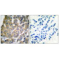
- Product NameGoat anti-COX2 / PTGS2
- Delivery Days Customer5
- Application Supplier NoteImmunofluorescence: Strong expression of the protein seen in the cytoplasm and vesicles of HepG2 and NIH3T3 cells. Recommended concentration: 10microg/ml. Flow Cytometry: Flow cytometric analysis of HeLa cells. Recommended concentration: 10ug/ml. Additional validation: This antibody has been successfully used in the following paper: Sikorski et al. (2018) PMID: 30377371.
- ApplicationsFlow Cytometry, ImmunoFluorescence, Western Blot, ELISA
- Applications SupplierPep-ELISA, WB, IF, FC
- CertificationResearch Use Only
- ClonalityPolyclonal
- Concentration0.5 mg/ml
- Gene ID5743
- Target namePTGS2
- Target descriptionprostaglandin-endoperoxide synthase 2
- Target synonymsCOX-2, COX2, GRIPGHS, PGG/HS, PGHS-2, PHS-2, hCox-2, prostaglandin G/H synthase 2, PGH synthase 2, PHS II, cyclooxygenase 2, cyclooxygenase 2b, prostaglandin H2 synthase 2, prostaglandin-endoperoxide synthase 2 (prostaglandin G/H synthase and cyclooxygenase)
- HostGoat
- Scientific DescriptionRefSeq number(s): NP_000954.1. Purification: Antigen affinity purified. Names and symbols: PTGS2; COX2; prostaglandin-endoperoxide synthase 2 (prostaglandin G/H synthase and cyclooxygenase); COX-2; PHS-2; PGG/HS; PGHS-2; hCox-2; prostaglandin G/H synthase and cyclooxygenase; GRIPGHS; cyclooxygenase 2b
- ReactivityBovine, Canine, Human, Mouse, Porcine
- Reactivity SupplierHuman, Dog, Pig, Cow
- Storage Instruction-20°C
- UNSPSC12352203
References
- Sikorski K, Mehta A, Inngjerdingen M, et al. A high-throughput pipeline for validation of antibodies. Nat Methods. 2018,15(11):909-912. doi: 10.1038/s41592-018-0179-8Read this paper
Related products
Product group Antibodies
Anti-Cox2 AntibodyA35580
ApplicationsImmunoFluorescence, Western Blot, ImmunoHistoChemistry
ReactivityHuman, Mouse, Rat
- SizePrice
Product group Antibodies
Anti-Cox2 Antibody144-62360
ApplicationsImmunoFluorescence, ImmunoPrecipitation, Western Blot, ImmunoHistoChemistry
ReactivityHuman, Mouse, Rat
TargetPTGS2
- SizePrice

Product group Antibodies
Anti-COX2/Cyclooxygenase 2/PTGS2 Picoband(r) AntibodyA00084-2-CARRIER-FREE
ApplicationsFlow Cytometry, Western Blot, ELISA, ImmunoHistoChemistry
ReactivityHuman, Mouse
TargetPTGS2
- SizePrice

Product group Antibodies
References
Cyclooxygenase 2 Polyclonal AntibodyBS-10411R
ApplicationsImmunoFluorescence, Western Blot, ELISA, ImmunoCytoChemistry, ImmunoHistoChemistry, ImmunoHistoChemistry Frozen, ImmunoHistoChemistry Paraffin
TargetPTGS2
- SizePrice
Product group Antibodies
PTGS2 Monoclonal AntibodyCSB-MA000320
ApplicationsELISA, ImmunoHistoChemistry
ReactivityHuman, Mouse, Rat
TargetPTGS2
- SizePrice
Product group Antibodies
PTGS2 Polyclonal AntibodyCAC15817
ApplicationsWestern Blot, ELISA
TargetPTGS2
- SizePrice
![Untreated (–) and treated (+) THP-1 whole cell extract (30 μg) were separated by 7.5% SDS-PAGE, and the membrane was blotted with COX2 antibody [C3], C-term (GTX100656) diluted at 1:500. The HRP-conjugated anti-rabbit IgG antibody (GTX213110-01) was used to detect the primary antibody, and the signal was developed with Trident ECL plus-Enhanced.](https://www.genetex.com/upload/website/prouct_img/normal/GTX100656/GTX100656_43222_20230203_WB_treatment_PMA_LPS_23020621_417.webp)
Product group Antibodies
COX2 antibody [C3], C-termGTX100656
ApplicationsWestern Blot, ImmunoHistoChemistry, ImmunoHistoChemistry Paraffin
ReactivityHuman, Mouse, Rat
TargetPTGS2
- SizePrice

Product group Antibodies
Anti-PTGS2 AntibodyHPA001335
ApplicationsImmunoCytoChemistry, ImmunoHistoChemistry
ReactivityHuman
TargetPTGS2
- SizePrice
Product group Antibodies
anti-COX-2 (human), Rabbit Monoclonal (RM348)REV-31-1234-00
ApplicationsWestern Blot, ImmunoHistoChemistry
ReactivityHuman
TargetPTGS2
- SizePrice