


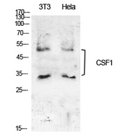
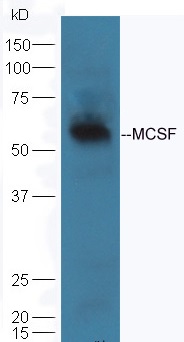
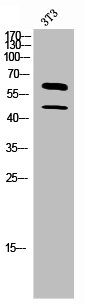
Figure 1. Western blot analysis of CSF1 using anti-CSF1 antibody (A00620-3). Electrophoresis was performed on a 5-20% SDS-PAGE gel at 70V (Stacking gel) / 90V (Resolving gel) for 2-3 hours. The sample well of each lane was loaded with 50ug of sample under reducing conditions. Lane 1: human placenta tissue lysates, Lane 2: human 293T whole cell lysate. After Electrophoresis, proteins were transferred to a Nitrocellulose membrane at 150mA for 50-90 minutes. Blocked the membrane with 5% Non-fat Milk/ TBS for 1.5 hour at RT. The membrane was incubated with rabbit anti-CSF1 antigen affinity purified polyclonal antibody (Catalog # A00620-3) at 0.5 microg/mL overnight at 4°C, then washed with TBS-0.1%Tween 3 times with 5 minutes each and probed with a goat anti-rabbit IgG-HRP secondary antibody at a dilution of 1:10000 for 1.5 hour at RT. The signal is developed using an Enhanced Chemiluminescent detection (ECL) kit (Catalog # EK1002) with Tanon 5200 system. A specific band was detected for CSF1 at approximately 60KD. The expected band size for CSF1 is at 60KD.
Figure 1. Western blot analysis of CSF1 using anti-CSF1 antibody (A00620-3). Electrophoresis was performed on a 5-20% SDS-PAGE gel at 70V (Stacking gel) / 90V (Resolving gel) for 2-3 hours. The sample well of each lane was loaded with 50ug of sample under reducing conditions. Lane 1: human placenta tissue lysates, Lane 2: human 293T whole cell lysate. After Electrophoresis, proteins were transferred to a Nitrocellulose membrane at 150mA for 50-90 minutes. Blocked the membrane with 5% Non-fat Milk/ TBS for 1.5 hour at RT. The membrane was incubated with rabbit anti-CSF1 antigen affinity purified polyclonal antibody (Catalog # A00620-3) at 0.5 microg/mL overnight at 4°C, then washed with TBS-0.1%Tween 3 times with 5 minutes each and probed with a goat anti-rabbit IgG-HRP secondary antibody at a dilution of 1:10000 for 1.5 hour at RT. The signal is developed using an Enhanced Chemiluminescent detection (ECL) kit (Catalog # EK1002) with Tanon 5200 system. A specific band was detected for CSF1 at approximately 60KD. The expected band size for CSF1 is at 60KD.
Anti-CSF1 Antibody Picoband(r)
A00620-3-CARRIER-FREE
ApplicationsWestern Blot, ELISA
Product group Antibodies
ReactivityHuman, Mouse
TargetCSF1
Overview
- SupplierBoster Bio
- Product NameAnti-CSF1 Antibody Picoband(r)
- Delivery Days Customer9
- ApplicationsWestern Blot, ELISA
- CertificationResearch Use Only
- ClonalityPolyclonal
- Concentration500 ug/ml
- Gene ID1435
- Target nameCSF1
- Target descriptioncolony stimulating factor 1
- Target synonymsCSF-1, MCSF, PG-M-CSF, macrophage colony-stimulating factor 1, CSF1-hERV-LTR71B fusion protein, colony stimulating factor 1 (macrophage), lanimostim, macrophage colony stimulating factor 1, proteoglycan macrophage colony-stimulating factor
- HostRabbit
- IsotypeIgG
- Protein IDP09603
- Protein NameMacrophage colony-stimulating factor 1
- Scientific DescriptionBoster Bio Anti-CSF1 Antibody Picoband® catalog # A00620-3. Tested in ELISA, WB applications. This antibody reacts with Human, Mouse. The brand Picoband indicates this is a premium antibody that guarantees superior quality, high affinity, and strong signals with minimal background in Western blot applications. Only our best-performing antibodies are designated as Picoband, ensuring unmatched performance.
- ReactivityHuman, Mouse
- Storage Instruction-20°C,2°C to 8°C
- UNSPSC12352203
Related products
Product group Antibodies
Anti-CSF1 AntibodyA101484
ApplicationsWestern Blot, ELISA
ReactivityHuman
- SizePrice
Product group Antibodies
Anti-CSF1 Antibody144-64248
ApplicationsWestern Blot, ImmunoHistoChemistry
ReactivityHuman
TargetCSF1
- SizePrice
Product group Antibodies

Anti-M-CSF [RX1], Mouse IgG1, kappaAB04366-1.1
ApplicationsNeutralisation/Blocking, Other Application
ReactivityHuman, Mouse
TargetCSF1
- SizePrice
Product group Antibodies

ApplicationsELISA
ReactivityHuman
TargetCSF1
- SizePrice
Product group Antibodies
CSF1 Polyclonal AntibodyBS-4910R
ApplicationsImmunoFluorescence, Western Blot, ELISA, ImmunoCytoChemistry, ImmunoHistoChemistry, ImmunoHistoChemistry Frozen, ImmunoHistoChemistry Paraffin
ReactivityBovine, Canine, Human, Mouse, Porcine, Rabbit, Rat
TargetCSF1
- SizePrice
Product group Antibodies
CSF1 AntibodyCSB-PA006115
ApplicationsWestern Blot, ELISA
ReactivityHuman
TargetCSF1
- SizePrice
Product group Antibodies
ApplicationsImmunoPrecipitation, Western Blot, ImmunoCytoChemistry, ImmunoHistoChemistry
TargetCSF1
- SizePrice
Product group Antibodies
M-CSF antibodyGTX10674
ApplicationsWestern Blot, ELISA
ReactivityHuman
TargetCSF1
- SizePrice