

Anti-M-CSF [RX1], Mouse IgG1, kappa
AB04366-1.1
ApplicationsNeutralisation/Blocking, Other Application
Product group Antibodies
ReactivityHuman, Mouse
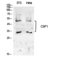
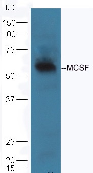
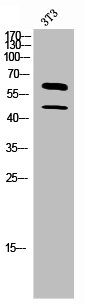
TargetCSF1
Overview
- SupplierAbsolute Antibody
- Product NameAnti-M-CSF [RX1], Mouse IgG1, kappa
- Delivery Days Customer9
- Application Supplier NoteThe binding properties of the original format of the antibody were analyzed using SPR analyses (Kd = 0.16 nM). The antibody could neutralize human M-CSF activity, as measured by a proliferation assay with M-NFS-60 cell line. Further, the Fab fragment of the antibody was able to neutralize human M-CSF activity, but with less potency. The humanized variant of the antibody was constructed (EP1913028B1).
- ApplicationsNeutralisation/Blocking, Other Application
- CertificationResearch Use Only
- ClonalityMonoclonal
- Clone IDRX1
- Gene ID1435
- Target nameCSF1
- Target descriptioncolony stimulating factor 1
- Target synonymsCSF-1, MCSF, PG-M-CSF, macrophage colony-stimulating factor 1, CSF1-hERV-LTR71B fusion protein, colony stimulating factor 1 (macrophage), lanimostim, macrophage colony stimulating factor 1, proteoglycan macrophage colony-stimulating factor
- HostMouse
- IsotypeIgG1
- Protein IDP09603
- Protein NameMacrophage colony-stimulating factor 1
- ReactivityHuman, Mouse
- Storage Instruction-20°C,2°C to 8°C
- UNSPSC41116161
Related products
Product group Antibodies
Anti-CSF1 AntibodyA101484
ApplicationsWestern Blot, ELISA
ReactivityHuman
- SizePrice

Product group Antibodies
Anti-CSF1 Antibody Picoband(r)A00620-3-CARRIER-FREE
ApplicationsWestern Blot, ELISA
ReactivityHuman, Mouse
TargetCSF1
- SizePrice
Product group Antibodies
Anti-CSF1 Antibody144-64248
ApplicationsWestern Blot, ImmunoHistoChemistry
ReactivityHuman
TargetCSF1
- SizePrice
Product group Antibodies

ApplicationsELISA
ReactivityHuman
TargetCSF1
- SizePrice
Product group Antibodies
CSF1 Polyclonal AntibodyBS-4910R
ApplicationsImmunoFluorescence, Western Blot, ELISA, ImmunoCytoChemistry, ImmunoHistoChemistry, ImmunoHistoChemistry Frozen, ImmunoHistoChemistry Paraffin
ReactivityBovine, Canine, Human, Mouse, Porcine, Rabbit, Rat
TargetCSF1
- SizePrice
Product group Antibodies
CSF1 AntibodyCSB-PA006115
ApplicationsWestern Blot, ELISA
ReactivityHuman
TargetCSF1
- SizePrice
Product group Antibodies
ApplicationsImmunoPrecipitation, Western Blot, ImmunoCytoChemistry, ImmunoHistoChemistry
TargetCSF1
- SizePrice
Product group Antibodies
M-CSF antibodyGTX10674
ApplicationsWestern Blot, ELISA
ReactivityHuman
TargetCSF1
- SizePrice