


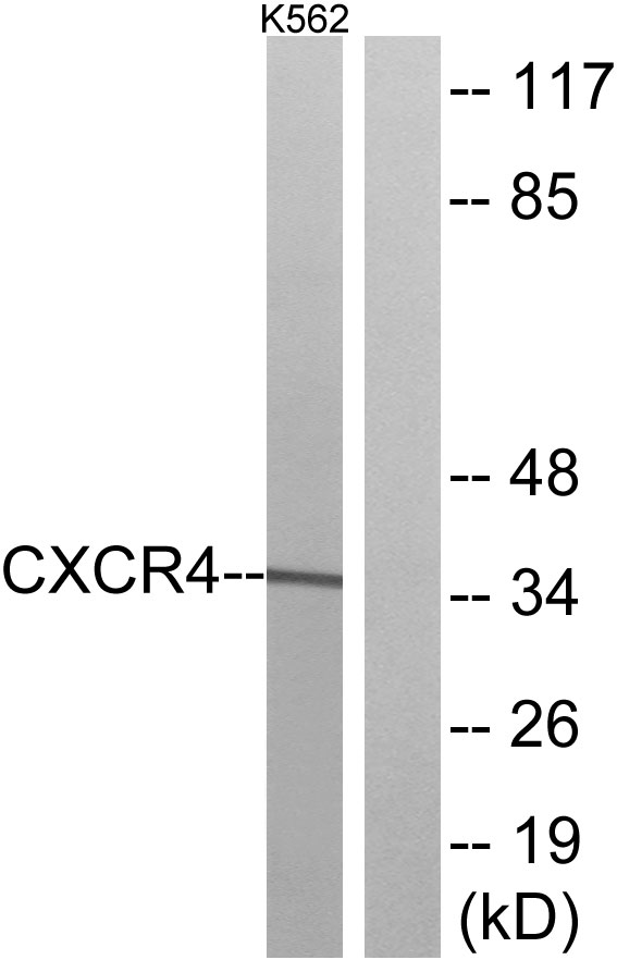
Figure 1. Western blot analysis of CXCR4 using anti-CXCR4 antibody (A00031-3). Electrophoresis was performed on a 5-20% SDS-PAGE gel at 70V (Stacking gel) / 90V (Resolving gel) for 2-3 hours. The sample well of each lane was loaded with 30ug of sample under reducing conditions. Lane 1: human Hela whole cell lysates, Lane 2: human HL-60 whole cell lysates, Lane 3: human SH-SY5Y whole cell lysates, Lane 4: human HEK293 whole cell lysates. After Electrophoresis, proteins were transferred to a Nitrocellulose membrane at 150mA for 50-90 minutes. Blocked the membrane with 5% Non-fat Milk/ TBS for 1.5 hour at RT. The membrane was incubated with rabbit anti-CXCR4 antigen affinity purified polyclonal antibody (Catalog # A00031-3) at 0.5 microg/mL overnight at 4°C, then washed with TBS-0.1%Tween 3 times with 5 minutes each and probed with a goat anti-rabbit IgG-HRP secondary antibody at a dilution of 1:5000 for 1.5 hour at RT. The signal is developed using an Enhanced Chemiluminescent detection (ECL) kit (Catalog # EK1002) with Tanon 5200 system. A specific band was detected for CXCR4 at approximately 60KD. The expected band size for CXCR4 is at 60KD.
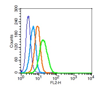
. Overlay histogram showing U937 cells stained with A00031-3 (Blue line).To facilitate intracellular staining, cells were fixed with 4% paraformaldehyde and permeabilized with permeabilization buffer. The cells were blocked with 10% normal goat serum. And then incubated with rabbit anti-CXCR4 Antibody (A00031-3, 1microg/1x106 cells) for 30 min at 20°C. DyLight®488 conjugated goat anti-rabbit IgG (BA1127, 5-10microg/1x106 cells) was used as secondary antibody for 30 minutes at 20°C. Isotype control antibody (Green line) was rabbit IgG (1microg/1x106) used under the same conditions. Unlabelled sample without incubation with primary antibody and secondary antibody (Red line) was used as a blank control.")
Figure 1. Western blot analysis of CXCR4 using anti-CXCR4 antibody (A00031-3). Electrophoresis was performed on a 5-20% SDS-PAGE gel at 70V (Stacking gel) / 90V (Resolving gel) for 2-3 hours. The sample well of each lane was loaded with 30ug of sample under reducing conditions. Lane 1: human Hela whole cell lysates, Lane 2: human HL-60 whole cell lysates, Lane 3: human SH-SY5Y whole cell lysates, Lane 4: human HEK293 whole cell lysates. After Electrophoresis, proteins were transferred to a Nitrocellulose membrane at 150mA for 50-90 minutes. Blocked the membrane with 5% Non-fat Milk/ TBS for 1.5 hour at RT. The membrane was incubated with rabbit anti-CXCR4 antigen affinity purified polyclonal antibody (Catalog # A00031-3) at 0.5 microg/mL overnight at 4°C, then washed with TBS-0.1%Tween 3 times with 5 minutes each and probed with a goat anti-rabbit IgG-HRP secondary antibody at a dilution of 1:5000 for 1.5 hour at RT. The signal is developed using an Enhanced Chemiluminescent detection (ECL) kit (Catalog # EK1002) with Tanon 5200 system. A specific band was detected for CXCR4 at approximately 60KD. The expected band size for CXCR4 is at 60KD.
Anti-CXCR4 Antibody Picoband(r)
A00031-3
ApplicationsFlow Cytometry, Western Blot, ELISA
Product group Antibodies
ReactivityHuman
TargetCXCR4
Overview
- SupplierBoster Bio
- Product NameAnti-CXCR4 Antibody Picoband(r)
- Delivery Days Customer9
- Application Supplier NoteTested Species: In-house tested species with positive results. Other applications have not been tested. Optimal dilutions should be determined by end users.
- ApplicationsFlow Cytometry, Western Blot, ELISA
- CertificationResearch Use Only
- ClonalityPolyclonal
- Concentration500 ug/ml
- Gene ID7852
- Target nameCXCR4
- Target descriptionC-X-C motif chemokine receptor 4
- Target synonymsCD184, D2S201E, FB22, HM89, HSY3RR, LAP-3, LAP3, LCR1, LESTR, NPY3R, NPYR, NPYRL, NPYY3R, WHIM, WHIMS, WHIMS1, C-X-C chemokine receptor type 4, CD184 antigen, LPS-associated protein 3, SDF-1 receptor, chemokine (C-X-C motif) receptor 4, fusin, leukocyte-derived seven transmembrane domain receptor, lipopolysaccharide-associated protein 3, neuropeptide Y receptor Y3, neuropeptide Y3 receptor, seven transmembrane helix receptor, seven-transmembrane-segment receptor, spleen, stromal cell-derived factor 1 receptor
- HostRabbit
- IsotypeIgG
- Protein IDP61073
- Protein NameC-X-C chemokine receptor type 4
- Scientific DescriptionBoster Bio Anti-CXCR4 Antibody Picoband® catalog # A00031-3. Tested in ELISA, Flow Cytometry, WB applications. This antibody reacts with Human. The brand Picoband indicates this is a premium antibody that guarantees superior quality, high affinity, and strong signals with minimal background in Western blot applications. Only our best-performing antibodies are designated as Picoband, ensuring unmatched performance.
- ReactivityHuman
- Storage Instruction-20°C,2°C to 8°C
- UNSPSC12352203
Related products
Product group Antibodies
Cxcr4 Polyclonal AntibodyCAC07015
ApplicationsImmunoFluorescence, Western Blot, ELISA, ImmunoHistoChemistry
ReactivityMouse
TargetCXCR4
- SizePrice
Product group Antibodies
References
CXCR4 Polyclonal AntibodyBS-1011R
ApplicationsFlow Cytometry, ImmunoFluorescence, Western Blot, ELISA, ImmunoCytoChemistry, ImmunoHistoChemistry, ImmunoHistoChemistry Frozen, ImmunoHistoChemistry Paraffin
ReactivityHuman, Mouse, Rat
TargetCXCR4
- SizePrice
Product group Antibodies
Anti-CXCR4 Antibody144-01303
ApplicationsWestern Blot
ReactivityHuman, Mouse
TargetCXCR4
- SizePrice
Product group Antibodies
Anti-CXCR4 AntibodyA96074
ApplicationsImmunoFluorescence, Western Blot, ELISA
ReactivityHuman, Mouse, Rat
- SizePrice
Product group Antibodies

Anti-CXCR4 [I-3859 ]Ab02396-1.1
ApplicationsFlow Cytometry, ImmunoPrecipitation, ELISA, ImmunoHistoChemistry
ReactivityHuman
TargetCXCR4
- SizePrice

Product group Antibodies
CXCR4 antibody [HL2612]GTX639064
ApplicationsImmunoFluorescence, Western Blot, ImmunoCytoChemistry
ReactivityCanine, Human, Mouse
TargetCXCR4
- SizePrice
Product group Antibodies

CXCR4 Antibody (Ser339)LS-C769147
ApplicationsImmunoFluorescence, Western Blot, ELISA, ImmunoHistoChemistry, ImmunoHistoChemistry Paraffin
ReactivityHuman, Monkey, Mouse, Rat
TargetCXCR4
- SizePrice