


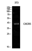
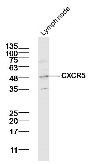
Figure 1. Western blot analysis of CXCR5 using anti-CXCR5 antibody (A00663-2). Electrophoresis was performed on a 5-20% SDS-PAGE gel at 70V (Stacking gel) / 90V (Resolving gel) for 2-3 hours. The sample well of each lane was loaded with 30 ug of sample under reducing conditions. Lane 1: human Raji whole cell lysates, Lane 2: human MOLT-4 whole cell lysates, Lane 3: human Hela whole cell lysates, Lane 4: rat brain tissue lysates, Lane 5: mouse brain tissue lysates, Lane 6: mouse lung tissue lysates. After electrophoresis, proteins were transferred to a nitrocellulose membrane at 150 mA for 50-90 minutes. Blocked the membrane with 5% non-fat milk/TBS for 1.5 hour at RT. The membrane was incubated with rabbit anti-CXCR5 antigen affinity purified polyclonal antibody (Catalog # A00663-2) at 0.5 microg/mL overnight at 4°C, then washed with TBS-0.1%Tween 3 times with 5 minutes each and probed with a goat anti-rabbit IgG-HRP secondary antibody at a dilution of 1:5000 for 1.5 hour at RT. The signal is developed using an Enhanced Chemiluminescent detection (ECL) kit (Catalog # EK1002) with Tanon 5200 system. A specific band was detected for CXCR5 at approximately 50 kDa. The expected band size for CXCR5 is at 42 kDa.
. Overlay histogram showing HEL cells stained with A00663-2 (Blue line). The cells were fixed with 4% paraformaldehyde and blocked with 10% normal goat serum. And then incubated with rabbit anti-CXCR5 Antibody (A00663-2, 1 microg/1x106 cells) for 30 min at 20°C. DyLight®488 conjugated goat anti-rabbit IgG (BA1127, 5-10 microg/1x106 cells) was used as secondary antibody for 30 minutes at 20°C. Isotype control antibody (Green line) was rabbit IgG (1 microg/1x106) used under the same conditions. Unlabelled sample without incubation with primary antibody and secondary antibody (Red line) was used as a blank control.")
Figure 1. Western blot analysis of CXCR5 using anti-CXCR5 antibody (A00663-2). Electrophoresis was performed on a 5-20% SDS-PAGE gel at 70V (Stacking gel) / 90V (Resolving gel) for 2-3 hours. The sample well of each lane was loaded with 30 ug of sample under reducing conditions. Lane 1: human Raji whole cell lysates, Lane 2: human MOLT-4 whole cell lysates, Lane 3: human Hela whole cell lysates, Lane 4: rat brain tissue lysates, Lane 5: mouse brain tissue lysates, Lane 6: mouse lung tissue lysates. After electrophoresis, proteins were transferred to a nitrocellulose membrane at 150 mA for 50-90 minutes. Blocked the membrane with 5% non-fat milk/TBS for 1.5 hour at RT. The membrane was incubated with rabbit anti-CXCR5 antigen affinity purified polyclonal antibody (Catalog # A00663-2) at 0.5 microg/mL overnight at 4°C, then washed with TBS-0.1%Tween 3 times with 5 minutes each and probed with a goat anti-rabbit IgG-HRP secondary antibody at a dilution of 1:5000 for 1.5 hour at RT. The signal is developed using an Enhanced Chemiluminescent detection (ECL) kit (Catalog # EK1002) with Tanon 5200 system. A specific band was detected for CXCR5 at approximately 50 kDa. The expected band size for CXCR5 is at 42 kDa.
Anti-CXCR5 Antibody Picoband(r)
A00663-2-CARRIER-FREE
ApplicationsFlow Cytometry, Western Blot, ELISA
Product group Antibodies
ReactivityHuman, Mouse, Rat
TargetCXCR5
Overview
- SupplierBoster Bio
- Product NameAnti-CXCR5 Antibody Picoband(r)
- Delivery Days Customer9
- ApplicationsFlow Cytometry, Western Blot, ELISA
- CertificationResearch Use Only
- ClonalityPolyclonal
- Concentration500 ug/ml
- Gene ID643
- Target nameCXCR5
- Target descriptionC-X-C motif chemokine receptor 5
- Target synonymsBLR1, CD185, MDR15, C-X-C chemokine receptor type 5, Burkitt lymphoma receptor 1, GTP binding protein (chemokine (C-X-C motif) receptor 5), Burkitt lymphoma receptor 1, GTP-binding protein, CXC-R5, CXCR-5, MDR-15, chemokine (C-X-C motif) receptor 5, monocyte-derived receptor 15
- HostRabbit
- IsotypeIgG
- Protein IDP32302
- Protein NameC-X-C chemokine receptor type 5
- Scientific DescriptionBoster Bio Anti-CXCR5 Antibody Picoband® catalog # A00663-2. Tested in ELISA, Flow Cytometry, WB applications. This antibody reacts with Human, Mouse, Rat. The brand Picoband indicates this is a premium antibody that guarantees superior quality, high affinity, and strong signals with minimal background in Western blot applications. Only our best-performing antibodies are designated as Picoband, ensuring unmatched performance.
- ReactivityHuman, Mouse, Rat
- Storage Instruction-20°C,2°C to 8°C
- UNSPSC12352203
Related products
Product group Antibodies
CXCR5 AntibodyCSB-PA006255LA01HU
ApplicationsImmunoFluorescence, ELISA, ImmunoHistoChemistry
ReactivityHuman
TargetCXCR5
- SizePrice
Product group Antibodies
Anti-CXCR5 AntibodyA101124
ApplicationsWestern Blot, ELISA
ReactivityHuman
- SizePrice
Product group Antibodies

CXCR5 AntibodyLS-C831029
ApplicationsELISA, ImmunoHistoChemistry
ReactivityHuman
TargetCXCR5
- SizePrice
Product group Antibodies
References
CXCR5 Polyclonal AntibodyBS-3598R
ApplicationsWestern Blot, ELISA, ImmunoHistoChemistry, ImmunoHistoChemistry Frozen, ImmunoHistoChemistry Paraffin
ReactivityBovine, Human, Mouse, Porcine, Rabbit, Rat
TargetCXCR5
- SizePrice
![Boiled and unboiled Raji whole cell and membrane extracts (30 μg) were separated by 10% SDS-PAGE, and the membrane was blotted with CXCR5 antibody [HL3568] (GTX641474) diluted at 1:1000. The HRP-conjugated anti-rabbit IgG antibody (GTX213110-01) was used to detect the primary antibody.](https://www.genetex.com/upload/website/prouct_img/normal/GTX641474/GTX641474_T-45621_20241213_WB_Fraction_ub_24121918_103.webp)
Product group Antibodies
CXCR5 antibody [HL3568]GTX641474
ApplicationsWestern Blot
ReactivityHuman
TargetCXCR5
- SizePrice
Product group Antibodies
Anti-CXCR5 (N-term) Antibody102-23722
ApplicationsWestern Blot
TargetCXCR5
- SizePrice