


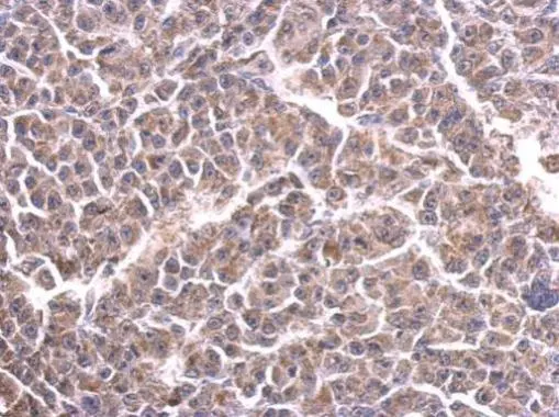
Immunohistochemical staining of human skeletal muscle shows moderate granular positivity in cytoplasm in myocytes.

Immunohistochemical staining of human skeletal muscle shows moderate granular positivity in cytoplasm in myocytes.
Anti-CYC1 Antibody
HPA001247
ApplicationsWestern Blot, ImmunoCytoChemistry, ImmunoHistoChemistry
Product group Antibodies
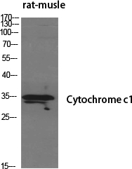
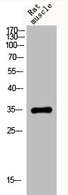
ReactivityHuman, Mouse, Rat
TargetCYC1
Overview
- SupplierAtlas Antibodies
- Product NameAnti-CYC1 Antibody
- Delivery Days Customer4
- ApplicationsWestern Blot, ImmunoCytoChemistry, ImmunoHistoChemistry
- CertificationResearch Use Only
- ClonalityPolyclonal
- ConjugateUnconjugated
- Gene ID1537
- Target nameCYC1
- Target descriptioncytochrome c1
- Target synonymsMC3DN6, UQCR4, cytochrome c1, heme protein, mitochondrial, complex III subunit 4, complex III subunit IV, cytochrome b-c1 complex subunit 4, cytochrome c-1, ubiquinol-cytochrome-c reductase complex cytochrome c1 subunit
- HostRabbit
- IsotypeIgG
- Protein IDP08574
- Protein NameCytochrome c1, heme protein, mitochondrial
- Scientific DescriptionRecombinant Protein Epitope Signature Tag (PrEST) antigen sequence
- ReactivityHuman, Mouse, Rat
- Storage Instruction-20°C,2°C to 8°C
- UNSPSC41116161
Datasheet
MSDS
Related products
Product group Antibodies
Anti-CYC1 AntibodyA99447
ApplicationsWestern Blot, ELISA
ReactivityHuman, Mouse
- SizePrice
Product group Antibodies
Anti-Cytochrome C Antibody130-10033
ApplicationsELISA
ReactivityHuman
TargetCYC1
- SizePrice

Product group Antibodies
Anti-CYC1 Antibody Picoband(r)A02958-2-CARRIER-FREE
ApplicationsFlow Cytometry, ImmunoFluorescence, Western Blot, ELISA, ImmunoCytoChemistry
ReactivityHuman
TargetCYC1
- SizePrice
Product group Antibodies
CYC1 Polyclonal AntibodyCAC13837
ApplicationsImmunoFluorescence, Western Blot, ELISA, ImmunoHistoChemistry
TargetCYC1
- SizePrice
Product group Antibodies
CYC1 AntibodyCSB-PA002014
ApplicationsWestern Blot, ELISA
ReactivityHuman, Mouse
TargetCYC1
- SizePrice
Product group Antibodies
CYC1 antibodyGTX101717
ApplicationsWestern Blot, ImmunoHistoChemistry, ImmunoHistoChemistry Paraffin
ReactivityHuman, Mouse
TargetCYC1
- SizePrice
Product group Antibodies

ApplicationsELISA
ReactivityHuman
TargetCYC1
- SizePrice