


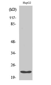
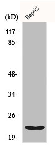
Figure 1. Western blot analysis of Cyclophilin F using anti-Cyclophilin F antibody (M02803). Electrophoresis was performed on a 5-20% SDS-PAGE gel at 70V (Stacking gel) / 90V (Resolving gel) for 2-3 hours. The sample well of each lane was loaded with 30 ug of sample under reducing conditions. Lane 1: human Hela whole cell lysates, Lane 2: human HEL whole cell lysates, Lane 3: human PC-3 whole cell lysates, Lane 4: human HepG2 whole cell lysates, Lane 5: rat PC-12 whole cell lysates, Lane 6: rat C6 whole cell lysates, Lane 7: mouse RAW264.7 whole cell lysates, Lane 8: mouse Neuro-2a whole cell lysates. After electrophoresis, proteins were transferred to a nitrocellulose membrane at 150 mA for 50-90 minutes. Blocked the membrane with 5% non-fat milk/TBS for 1.5 hour at RT. The membrane was incubated with rabbit anti-Cyclophilin F antigen affinity purified monoclonal antibody (Catalog # M02803) at 1:500 overnight at 4°C, then washed with TBS-0.1%Tween 3 times with 5 minutes each and probed with a goat anti-rabbit IgG-HRP secondary antibody at a dilution of 1:5000 for 1.5 hour at RT. The signal is developed using an Enhanced Chemiluminescent detection (ECL) kit (Catalog # EK1002) with Tanon 5200 system. A specific band was detected for Cyclophilin F at approximately 18 kDa. The expected band size for Cyclophilin F is at 22 kDa.
Figure 1. Western blot analysis of Cyclophilin F using anti-Cyclophilin F antibody (M02803). Electrophoresis was performed on a 5-20% SDS-PAGE gel at 70V (Stacking gel) / 90V (Resolving gel) for 2-3 hours. The sample well of each lane was loaded with 30 ug of sample under reducing conditions. Lane 1: human Hela whole cell lysates, Lane 2: human HEL whole cell lysates, Lane 3: human PC-3 whole cell lysates, Lane 4: human HepG2 whole cell lysates, Lane 5: rat PC-12 whole cell lysates, Lane 6: rat C6 whole cell lysates, Lane 7: mouse RAW264.7 whole cell lysates, Lane 8: mouse Neuro-2a whole cell lysates. After electrophoresis, proteins were transferred to a nitrocellulose membrane at 150 mA for 50-90 minutes. Blocked the membrane with 5% non-fat milk/TBS for 1.5 hour at RT. The membrane was incubated with rabbit anti-Cyclophilin F antigen affinity purified monoclonal antibody (Catalog # M02803) at 1:500 overnight at 4°C, then washed with TBS-0.1%Tween 3 times with 5 minutes each and probed with a goat anti-rabbit IgG-HRP secondary antibody at a dilution of 1:5000 for 1.5 hour at RT. The signal is developed using an Enhanced Chemiluminescent detection (ECL) kit (Catalog # EK1002) with Tanon 5200 system. A specific band was detected for Cyclophilin F at approximately 18 kDa. The expected band size for Cyclophilin F is at 22 kDa.
Anti-Cyclophilin F Rabbit Monoclonal Antibody
M02803
ApplicationsImmunoPrecipitation, Western Blot, ImmunoHistoChemistry
Product group Antibodies
ReactivityHuman, Mouse, Rat
TargetPPIF
Overview
- SupplierBoster Bio
- Product NameAnti-Cyclophilin F Rabbit Monoclonal Antibody
- Delivery Days Customer9
- ApplicationsImmunoPrecipitation, Western Blot, ImmunoHistoChemistry
- CertificationResearch Use Only
- ClonalityMonoclonal
- Clone IDACGO-16
- Gene ID10105
- Target namePPIF
- Target descriptionpeptidylprolyl isomerase F
- Target synonymsCYP3, CyP-M, Cyp-D, CypD, peptidyl-prolyl cis-trans isomerase F, mitochondrial, PPIase F, cyclophilin 3, cyclophilin D, cyclophilin F, mitochondrial cyclophilin, peptidyl-prolyl cis-trans isomerase, mitochondrial, rotamase F
- HostRabbit
- IsotypeIgG
- Protein IDP30405
- Protein NamePeptidyl-prolyl cis-trans isomerase F, mitochondrial
- Scientific DescriptionBoster Bio Anti-Cyclophilin F Rabbit Monoclonal Antibody catalog # M02803. Tested in WB, IHC, IP applications. This antibody reacts with Human, Mouse, Rat.
- ReactivityHuman, Mouse, Rat
- Storage Instruction-20°C
- UNSPSC12352203
Datasheet
MSDS
Related products
Product group Antibodies
Anti-PPIF Antibody144-03208
ApplicationsWestern Blot
ReactivityHuman, Mouse
TargetPPIF
- SizePrice
Product group Antibodies
ApplicationsImmunoFluorescence, Western Blot, ELISA, ImmunoHistoChemistry
ReactivityHuman, Mouse, Rat
- SizePrice
Product group Antibodies
Cyclophilin F Recombinant Antibody, AbBy Fluor-350 ConjugatedBSM-61600R-BF350
ApplicationsImmunoFluorescence, Western Blot
ReactivityHuman, Mouse, Rat
TargetPPIF
- SizePrice
Product group Antibodies
PPIF AntibodyCSB-PA001907
ApplicationsImmunoFluorescence, Western Blot, ELISA, ImmunoHistoChemistry
ReactivityHuman, Mouse, Rat
TargetPPIF
- SizePrice
Product group Antibodies
ApplicationsImmunoPrecipitation, Western Blot, ImmunoCytoChemistry, ImmunoHistoChemistry
ReactivityMouse, Rat
TargetPPIF
- SizePrice
Product group Antibodies

PPIF / Cyclophilin F AntibodyLS-C401561
ApplicationsWestern Blot, ELISA, ImmunoHistoChemistry
ReactivityHuman, Mouse, Rat
TargetPPIF
- SizePrice

Product group Antibodies
Anti-PPIF AntibodyHPA077416
ApplicationsImmunoCytoChemistry
ReactivityHuman
TargetPPIF
- SizePrice
Product group Antibodies
Cyclophilin F antibody [N1C3]GTX117951
ApplicationsImmunoFluorescence, Western Blot, ImmunoCytoChemistry, ImmunoHistoChemistry, ImmunoHistoChemistry Paraffin
ReactivityHuman, Mouse, Rat
TargetPPIF
- SizePrice