


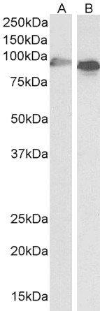
Figure 1. Western blot analysis of CYP1A1 using anti-CYP1A1 antibody (PB9544). Electrophoresis was performed on a 5-20% SDS-PAGE gel at 70V (Stacking gel) / 90V (Resolving gel) for 2-3 hours. The sample well of each lane was loaded with 50ug of sample under reducing conditions. Lane 1: Rat Lung Tissue Lysate Lane 2: Mouse Lung Tissue Lysate Lane 3: Human Placenta Tissue Lysate Lane 4: JURKAT Whole Cell Lysate After Electrophoresis, proteins were transferred to a Nitrocellulose membrane at 150mA for 50-90 minutes. Blocked the membrane with 5% Non-fat Milk/ TBS for 1.5 hour at RT. The membrane was incubated with rabbit anti-CYP1A1 antigen affinity purified polyclonal antibody (Catalog # PB9544) at 0.5 microg/mL overnight at 4°C, then washed with TBS-0.1%Tween 3 times with 5 minutes each and probed with a goat anti-rabbit IgG-HRP secondary antibody at a dilution of 1:10000 for 1.5 hour at RT. The signal is developed using an Enhanced Chemiluminescent detection (ECL) kit (Catalog # EK1002) with Tanon 5200 system. A specific band was detected for CYP1A1 at approximately 58KD. The expected band size for CYP1A1 is at 58KD.
. CYP1A1 was detected in paraffin-embedded section of Mouse Kidney Tissue. Heat mediated antigen retrieval was performed in citrate buffer (pH6, epitope retrieval solution) for 20 mins. The tissue section was blocked with 10% goat serum. The tissue section was then incubated with 1microg/ml rabbit anti-CYP1A1 Antibody (PB9544) overnight at 4°C. Biotinylated goat anti-rabbit IgG was used as secondary antibody and incubated for 30 minutes at 37°C. The tissue section was developed using Strepavidin-Biotin-Complex (SABC)(Catalog # SA1022) with DAB as the chromogen.")
. CYP1A1 was detected in paraffin-embedded section of Rat Kidney Tissue. Heat mediated antigen retrieval was performed in citrate buffer (pH6, epitope retrieval solution) for 20 mins. The tissue section was blocked with 10% goat serum. The tissue section was then incubated with 1microg/ml rabbit anti-CYP1A1 Antibody (PB9544) overnight at 4°C. Biotinylated goat anti-rabbit IgG was used as secondary antibody and incubated for 30 minutes at 37°C. The tissue section was developed using Strepavidin-Biotin-Complex (SABC)(Catalog # SA1022) with DAB as the chromogen.")
. CYP1A1 was detected in paraffin-embedded section of Human Mammary Cancer Tissue. Heat mediated antigen retrieval was performed in citrate buffer (pH6, epitope retrieval solution) for 20 mins. The tissue section was blocked with 10% goat serum. The tissue section was then incubated with 1microg/ml rabbit anti-CYP1A1 Antibody (PB9544) overnight at 4°C. Biotinylated goat anti-rabbit IgG was used as secondary antibody and incubated for 30 minutes at 37°C. The tissue section was developed using Strepavidin-Biotin-Complex (SABC)(Catalog # SA1022) with DAB as the chromogen.")
. CYP1A1 was detected in frozen section of Mouse Kidney Tissue. The tissue section was blocked with 10% goat serum. The tissue section was then incubated with 1microg/ml rabbit anti-CYP1A1 Antibody (PB9544) overnight at 4°C. Biotinylated goat anti-rabbit IgG was used as secondary antibody and incubated for 30 minutes at 37°C. The tissue section was developed using Strepavidin-Biotin-Complex (SABC)(Catalog # SA1022) with DAB as the chromogen.")
. CYP1A1 was detected in frozen section of Human Placenta Tissue. The tissue section was blocked with 10% goat serum. The tissue section was then incubated with 1microg/ml rabbit anti-CYP1A1 Antibody (PB9544) overnight at 4°C. Biotinylated goat anti-rabbit IgG was used as secondary antibody and incubated for 30 minutes at 37°C. The tissue section was developed using Strepavidin-Biotin-Complex (SABC)(Catalog # SA1022) with DAB as the chromogen.")
. Overlay histogram showing CACO-2 cells stained with PB9544 (Blue line). To facilitate intracellular staining, cells were fixed with 4% paraformaldehyde and permeabilized with permeabilization buffer. The cells were blocked with 10% normal goat serum. And then incubated with rabbit anti-CYP1A1 Antibody (PB9544,1microg/1x106 cells) for 30 min at 20°C. DyLight®488 conjugated goat anti-rabbit IgG (BA1127, 5-10microg/1x106 cells) was used as secondary antibody for 30 minutes at 20°C. Isotype control antibody (Green line) was rabbit IgG (1microg/1x106) used under the same conditions. Unlabelled sample without incubation with primary antibody and secondary antibody (Red line) was used as a blank control.")
. Overlay histogram showing Hela cells stained with PB9544 (Blue line). To facilitate intracellular staining, cells were fixed with 4% paraformaldehyde and permeabilized with permeabilization buffer. The cells were blocked with 10% normal goat serum. And then incubated with rabbit anti-CYP1A1 Antibody (PB9544,1microg/1x106 cells) for 30 min at 20°C. DyLight®488 conjugated goat anti-rabbit IgG (BA1127, 5-10microg/1x106 cells) was used as secondary antibody for 30 minutes at 20°C. Isotype control antibody (Green line) was rabbit IgG (1microg/1x106) used under the same conditions. Unlabelled sample without incubation with primary antibody and secondary antibody (Red line) was used as a blank control.")
. Overlay histogram showing K562 cells stained with PB9544 (Blue line). To facilitate intracellular staining, cells were fixed with 4% paraformaldehyde and permeabilized with permeabilization buffer. The cells were blocked with 10% normal goat serum. And then incubated with rabbit anti-CYP1A1 Antibody (PB9544,1microg/1x106 cells) for 30 min at 20°C. DyLight®488 conjugated goat anti-rabbit IgG (BA1127, 5-10microg/1x106 cells) was used as secondary antibody for 30 minutes at 20°C. Isotype control antibody (Green line) was rabbit IgG (1microg/1x106) used under the same conditions. Unlabelled sample without incubation with primary antibody and secondary antibody (Red line) was used as a blank control.")
. CYP1A1 was detected in immunocytochemical section of Caco-2 cells. Enzyme antigen retrieval was performed using IHC enzyme antigen retrieval reagent (AR0022) for 15 mins. The cells were blocked with 10% goat serum. And then incubated with 2microg/mL rabbit anti-CYP1A1 Antibody (PB9544) overnight at 4°C. DyLight®488 Conjugated Goat Anti-Rabbit IgG (BA1127) was used as secondary antibody at 1:100 dilution and incubated for 30 minutes at 37°C. The section was counterstained with DAPI. Visualize using a fluorescence microscope and filter sets appropriate for the label used.")
Figure 1. Western blot analysis of CYP1A1 using anti-CYP1A1 antibody (PB9544). Electrophoresis was performed on a 5-20% SDS-PAGE gel at 70V (Stacking gel) / 90V (Resolving gel) for 2-3 hours. The sample well of each lane was loaded with 50ug of sample under reducing conditions. Lane 1: Rat Lung Tissue Lysate Lane 2: Mouse Lung Tissue Lysate Lane 3: Human Placenta Tissue Lysate Lane 4: JURKAT Whole Cell Lysate After Electrophoresis, proteins were transferred to a Nitrocellulose membrane at 150mA for 50-90 minutes. Blocked the membrane with 5% Non-fat Milk/ TBS for 1.5 hour at RT. The membrane was incubated with rabbit anti-CYP1A1 antigen affinity purified polyclonal antibody (Catalog # PB9544) at 0.5 microg/mL overnight at 4°C, then washed with TBS-0.1%Tween 3 times with 5 minutes each and probed with a goat anti-rabbit IgG-HRP secondary antibody at a dilution of 1:10000 for 1.5 hour at RT. The signal is developed using an Enhanced Chemiluminescent detection (ECL) kit (Catalog # EK1002) with Tanon 5200 system. A specific band was detected for CYP1A1 at approximately 58KD. The expected band size for CYP1A1 is at 58KD.
Anti-Cytochrome P450 1A1/CYP1A1 Antibody Picoband(r)
PB9544-CARRIER-FREE
ApplicationsFlow Cytometry, ImmunoFluorescence, Western Blot, ImmunoCytoChemistry, ImmunoHistoChemistry, ImmunoHistoChemistry Frozen
Product group Antibodies
ReactivityHuman, Mouse, Rat
TargetCYP1A1
Overview
- SupplierBoster Bio
- Product NameAnti-Cytochrome P450 1A1/CYP1A1 Antibody Picoband(r)
- Delivery Days Customer9
- Application Supplier NoteTested Species: In-house tested species with positive results. By Heat: Boiling the paraffin sections in 10mM citrate buffer, pH6.0, for 20mins is required for the staining of formalin/paraffin sections. Other applications have not been tested. Optimal dilutions should be determined by end users.
- ApplicationsFlow Cytometry, ImmunoFluorescence, Western Blot, ImmunoCytoChemistry, ImmunoHistoChemistry, ImmunoHistoChemistry Frozen
- CertificationResearch Use Only
- ClonalityPolyclonal
- Concentration500 ug/ml
- Gene ID1543
- Target nameCYP1A1
- Target descriptioncytochrome P450 family 1 subfamily A member 1
- Target synonymsAHH, AHRR, CP11, CYP1, CYPIA1, P1-450, P450-C, P450DX, cytochrome P450 1A1, aryl hydrocarbon hydroxylase, cytochrome P1-450, dioxin-inducible, cytochrome P450 form 6, cytochrome P450, family 1, subfamily A, polypeptide 1, cytochrome P450, subfamily I (aromatic compound-inducible), polypeptide 1, cytochrome P450-C, cytochrome P450-P1, flavoprotein-linked monooxygenase, hydroperoxy icosatetraenoate dehydratase, xenobiotic monooxygenase
- HostRabbit
- IsotypeIgG
- Protein IDP04798
- Protein NameCytochrome P450 1A1
- Scientific DescriptionBoster Bio Anti-Cytochrome P450 1A1/CYP1A1 Antibody Picoband® catalog # PB9544. Tested in Flow Cytometry, IF, IHC, IHC-F, ICC, WB applications. This antibody reacts with Human, Mouse, Rat. The brand Picoband indicates this is a premium antibody that guarantees superior quality, high affinity, and strong signals with minimal background in Western blot applications. Only our best-performing antibodies are designated as Picoband, ensuring unmatched performance.
- ReactivityHuman, Mouse, Rat
- Storage Instruction-20°C,2°C to 8°C
- UNSPSC12352203
Related products
Product group Antibodies
Anti-CYP1A1 AntibodyA82489
ApplicationsWestern Blot, ELISA
ReactivityHuman
- SizePrice
Product group Antibodies
Anti-CYP1A1 Antibody144-02159
ApplicationsWestern Blot, ImmunoHistoChemistry
ReactivityHuman, Mouse, Rat
TargetCYP1A1
- SizePrice
Product group Antibodies

CYP1A1 AntibodyLS-C765943
ApplicationsWestern Blot, ELISA, ImmunoHistoChemistry
ReactivityHuman, Mouse, Rat
TargetCYP1A1
- SizePrice
Product group Antibodies
CYP1A1 (10G3) Monoclonal AntibodyBSM-51210M
ApplicationsWestern Blot, ELISA
ReactivityHuman
TargetCYP1A1
- SizePrice
Product group Antibodies
CYP1A1/CYP1A2 AntibodyCSB-PA001929
ApplicationsImmunoFluorescence, Western Blot, ELISA, ImmunoHistoChemistry
ReactivityHuman, Monkey, Mouse, Rat
TargetCYP1A1
- SizePrice
Product group Antibodies

ApplicationsWestern Blot, ELISA
ReactivityHuman
TargetCYP1A1
- SizePrice
Product group Antibodies
ApplicationsImmunoPrecipitation, Western Blot, ImmunoCytoChemistry, ImmunoHistoChemistry
ReactivityMouse, Rat
TargetCYP1A1
- SizePrice
Product group Antibodies
References
CYP1A1 antibodyGTX55582
ApplicationsWestern Blot, ImmunoHistoChemistry, ImmunoHistoChemistry Paraffin
ReactivityHuman, Mouse, Rat
TargetCYP1A1
- SizePrice
Product group Antibodies
Anti-CYP1A1 AntibodyCAB2159
ApplicationsImmunoFluorescence, Western Blot, ELISA, ImmunoCytoChemistry
ReactivityHuman
TargetCYP1A1
- SizePrice