


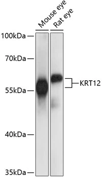
Figure 1. Western blot analysis of KRT12 using anti-KRT12 antibody (M05227). Electrophoresis was performed on a 5-20% SDS-PAGE gel at 70V (Stacking gel) / 90V (Resolving gel) for 2-3 hours. The sample well of each lane was loaded with 30 ug of sample under reducing conditions. Lane 1: rat eye tissue lysates, Lane 2: mouse eye tissue lysates. After electrophoresis, proteins were transferred to a nitrocellulose membrane at 150 mA for 50-90 minutes. Blocked the membrane with 5% non-fat milk/TBS for 1.5 hour at RT. The membrane was incubated with rabbit anti-KRT12 antigen affinity purified monoclonal antibody (Catalog # M05227) at 1:500 overnight at 4°C, then washed with TBS-0.1%Tween 3 times with 5 minutes each and probed with a goat anti-rabbit IgG-HRP secondary antibody at a dilution of 1:500 for 1.5 hour at RT. The signal is developed using an Enhanced Chemiluminescent detection (ECL) kit (Catalog # EK1002) with Tanon 5200 system. A specific band was detected for KRT12 at approximately 54 kDa. The expected band size for KRT12 is at 54 kDa.
Figure 1. Western blot analysis of KRT12 using anti-KRT12 antibody (M05227). Electrophoresis was performed on a 5-20% SDS-PAGE gel at 70V (Stacking gel) / 90V (Resolving gel) for 2-3 hours. The sample well of each lane was loaded with 30 ug of sample under reducing conditions. Lane 1: rat eye tissue lysates, Lane 2: mouse eye tissue lysates. After electrophoresis, proteins were transferred to a nitrocellulose membrane at 150 mA for 50-90 minutes. Blocked the membrane with 5% non-fat milk/TBS for 1.5 hour at RT. The membrane was incubated with rabbit anti-KRT12 antigen affinity purified monoclonal antibody (Catalog # M05227) at 1:500 overnight at 4°C, then washed with TBS-0.1%Tween 3 times with 5 minutes each and probed with a goat anti-rabbit IgG-HRP secondary antibody at a dilution of 1:500 for 1.5 hour at RT. The signal is developed using an Enhanced Chemiluminescent detection (ECL) kit (Catalog # EK1002) with Tanon 5200 system. A specific band was detected for KRT12 at approximately 54 kDa. The expected band size for KRT12 is at 54 kDa.
Anti-Cytokeratin 12 Rabbit Monoclonal Antibody
M05227
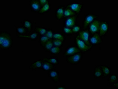
ApplicationsImmunoFluorescence, Western Blot, ImmunoCytoChemistry, ImmunoHistoChemistry
Product group Antibodies
ReactivityHuman, Mouse, Rat
TargetKRT12
Overview
- SupplierBoster Bio
- Product NameAnti-Cytokeratin 12 Rabbit Monoclonal Antibody
- Delivery Days Customer9
- ApplicationsImmunoFluorescence, Western Blot, ImmunoCytoChemistry, ImmunoHistoChemistry
- CertificationResearch Use Only
- ClonalityMonoclonal
- Clone ID26K31
- Gene ID3859
- Target nameKRT12
- Target descriptionkeratin 12
- Target synonymsK12, MECD1, keratin, type I cytoskeletal 12, CK-12, cytokeratin-12, keratin 12, type I
- HostRabbit
- IsotypeIgG
- Protein IDQ99456
- Protein NameKeratin, type I cytoskeletal 12
- Scientific DescriptionBoster Bio Anti-Cytokeratin 12 Rabbit Monoclonal Antibody catalog # M05227. Tested in WB, IHC, ICC/IF applications. This antibody reacts with Human, Mouse, Rat.
- ReactivityHuman, Mouse, Rat
- Storage Instruction-20°C
- UNSPSC12352203
Related products
Product group Antibodies
Anti-KRT12 Antibody144-60772
ApplicationsWestern Blot
ReactivityHuman, Mouse, Rat
TargetKRT12
- SizePrice
Product group Antibodies

KRT12 / CK12 / Cytokeratin 12 AntibodyLS-C749728
ApplicationsWestern Blot
ReactivityHuman, Mouse, Rat
TargetKRT12
- SizePrice
Product group Antibodies
CK12 Recombinant Antibody, AbBy Fluor-555 ConjugatedBSM-62422R-BF555
ApplicationsImmunoFluorescence, Western Blot
ReactivityHuman, Mouse, Rat
TargetKRT12
- SizePrice
Product group Antibodies
KRT12 AntibodyCSB-PA859510LA01HU
ApplicationsImmunoFluorescence, ELISA
ReactivityHuman
TargetKRT12
- SizePrice

Product group Antibodies
Anti-KRT12 AntibodyHPA055217
ApplicationsImmunoHistoChemistry
ReactivityHuman
TargetKRT12
- SizePrice
Product group Antibodies
Cytokeratin 12 antibodyGTX66286
ApplicationsWestern Blot
ReactivityHuman, Mouse, Rat
TargetKRT12
- SizePrice
Product group Antibodies
ApplicationsWestern Blot, ELISA
ReactivityMouse
TargetKRT12
- SizePrice