


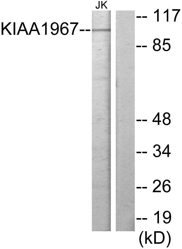
Figure 1. Western blot analysis of DBC-1/CCAR2 using anti-DBC-1/CCAR2 antibody (A03412-1). Electrophoresis was performed on a 5-20% SDS-PAGE gel at 70V (Stacking gel) / 90V (Resolving gel) for 2-3 hours. The sample well of each lane was loaded with 30 ug of sample under reducing conditions. Lane 1: human Jurkat whole cell lysates, Lane 2: monkey COS-7 whole cell lysates, Lane 3: human RT4 whole cell lysates, Lane 4: human K562 whole cell lysates, Lane 5: human HEL whole cell lysates, Lane 6: rat brain tissue lysates, Lane 7: mouse brain tissue lysates, Lane 8: mouse NIH/3T3 whole cell lysates. After electrophoresis, proteins were transferred to a nitrocellulose membrane at 150 mA for 50-90 minutes. Blocked the membrane with 5% non-fat milk/TBS for 1.5 hour at RT. The membrane was incubated with rabbit anti-DBC-1/CCAR2 antigen affinity purified polyclonal antibody (Catalog # A03412-1) at 0.25 microg/mL overnight at 4°C, then washed with TBS-0.1%Tween 3 times with 5 minutes each and probed with a goat anti-rabbit IgG-HRP secondary antibody at a dilution of 1:5000 for 1.5 hour at RT. The signal is developed using an Enhanced Chemiluminescent detection (ECL) kit (Catalog # EK1002) with Tanon 5200 system. A specific band was detected for DBC-1/CCAR2 at approximately 130 kDa. The expected band size for DBC-1/CCAR2 is at 130 kDa.
. DBC-1/CCAR2 was detected in a paraffin-embedded section of human gall bladder adenosquamous carcinoma tissue. Heat mediated antigen retrieval was performed in EDTA buffer (pH 8.0, epitope retrieval solution). The tissue section was blocked with 10% goat serum. The tissue section was then incubated with 2 microg/ml rabbit anti-DBC-1/CCAR2 Antibody (A03412-1) overnight at 4°C. Biotinylated goat anti-rabbit IgG was used as secondary antibody and incubated for 30 minutes at 37°C. The tissue section was developed using Strepavidin-Biotin-Complex (SABC) (Catalog # SA1022) with DAB as the chromogen.")
. DBC-1/CCAR2 was detected in a paraffin-embedded section of human liver cancer tissue. Heat mediated antigen retrieval was performed in EDTA buffer (pH 8.0, epitope retrieval solution). The tissue section was blocked with 10% goat serum. The tissue section was then incubated with 2 microg/ml rabbit anti-DBC-1/CCAR2 Antibody (A03412-1) overnight at 4°C. Biotinylated goat anti-rabbit IgG was used as secondary antibody and incubated for 30 minutes at 37°C. The tissue section was developed using Strepavidin-Biotin-Complex (SABC) (Catalog # SA1022) with DAB as the chromogen.")
. DBC-1/CCAR2 was detected in a paraffin-embedded section of human lung cancer tissue. Heat mediated antigen retrieval was performed in EDTA buffer (pH 8.0, epitope retrieval solution). The tissue section was blocked with 10% goat serum. The tissue section was then incubated with 2 microg/ml rabbit anti-DBC-1/CCAR2 Antibody (A03412-1) overnight at 4°C. Biotinylated goat anti-rabbit IgG was used as secondary antibody and incubated for 30 minutes at 37°C. The tissue section was developed using Strepavidin-Biotin-Complex (SABC) (Catalog # SA1022) with DAB as the chromogen.")
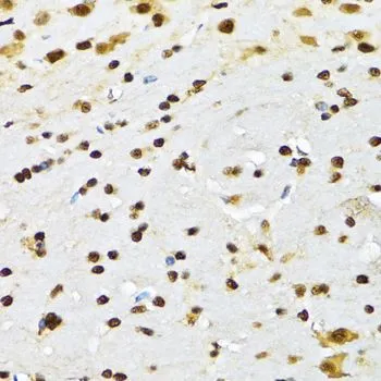
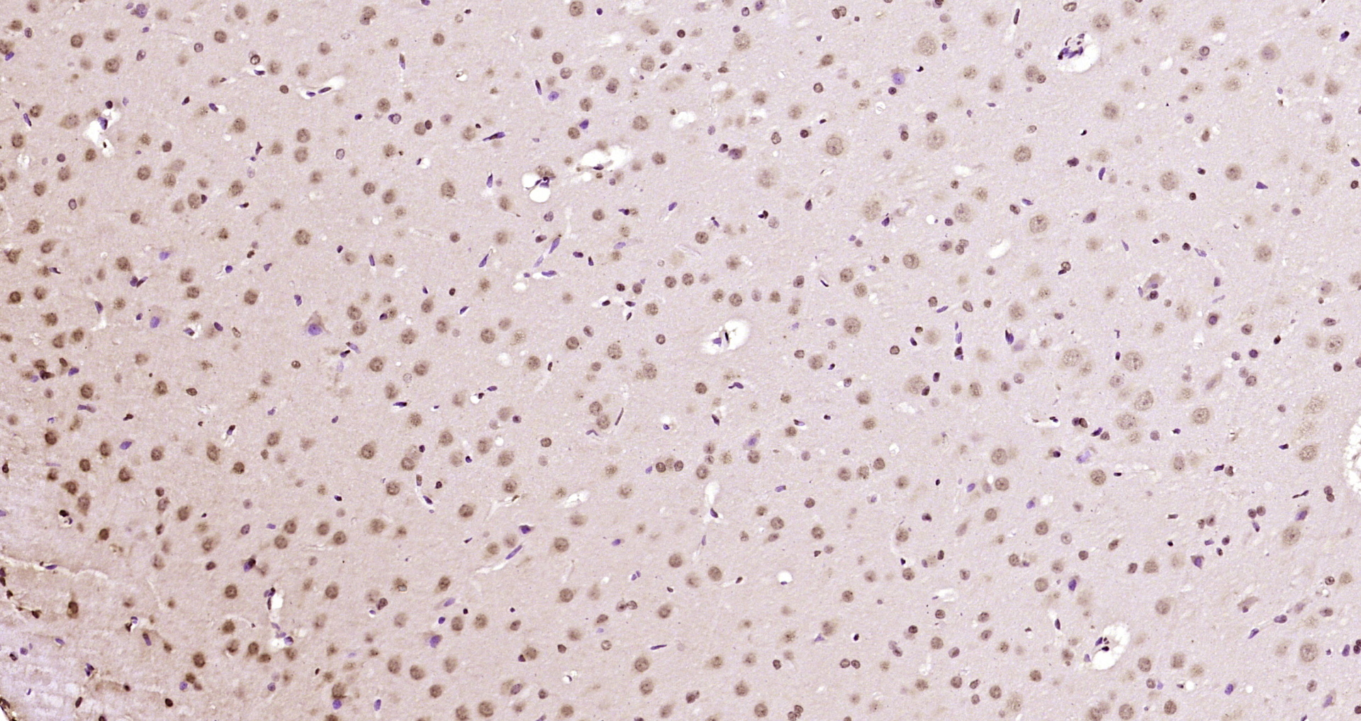
. DBC-1/CCAR2 was detected in a paraffin-embedded section of mouse brain tissue. Heat mediated antigen retrieval was performed in EDTA buffer (pH 8.0, epitope retrieval solution). The tissue section was blocked with 10% goat serum. The tissue section was then incubated with 2 microg/ml rabbit anti-DBC-1/CCAR2 Antibody (A03412-1) overnight at 4°C. Biotinylated goat anti-rabbit IgG was used as secondary antibody and incubated for 30 minutes at 37°C. The tissue section was developed using Strepavidin-Biotin-Complex (SABC) (Catalog # SA1022) with DAB as the chromogen.")
. DBC-1/CCAR2 was detected in a paraffin-embedded section of rat brain tissue. Heat mediated antigen retrieval was performed in EDTA buffer (pH 8.0, epitope retrieval solution). The tissue section was blocked with 10% goat serum. The tissue section was then incubated with 2 microg/ml rabbit anti-DBC-1/CCAR2 Antibody (A03412-1) overnight at 4°C. Biotinylated goat anti-rabbit IgG was used as secondary antibody and incubated for 30 minutes at 37°C. The tissue section was developed using Strepavidin-Biotin-Complex (SABC) (Catalog # SA1022) with DAB as the chromogen.")
. DBC-1/CCAR2 was detected in a paraffin-embedded section of human laryngeal carcinoma tissue. Heat mediated antigen retrieval was performed in EDTA buffer (pH 8.0, epitope retrieval solution). The tissue section was blocked with 10% goat serum. The tissue section was then incubated with 2 microg/ml rabbit anti-DBC-1/CCAR2 Antibody (A03412-1) overnight at 4°C. Biotinylated goat anti-rabbit IgG was used as secondary antibody and incubated for 30 minutes at 37°C. The tissue section was developed using Strepavidin-Biotin-Complex (SABC) (Catalog # SA1022) with DAB as the chromogen.")
. DBC-1/CCAR2 was detected in a paraffin-embedded section of human pancreatic carcinoma tissue. Heat mediated antigen retrieval was performed in EDTA buffer (pH 8.0, epitope retrieval solution). The tissue section was blocked with 10% goat serum. The tissue section was then incubated with 2 microg/ml rabbit anti-DBC-1/CCAR2 Antibody (A03412-1) overnight at 4°C. Biotinylated goat anti-rabbit IgG was used as secondary antibody and incubated for 30 minutes at 37°C. The tissue section was developed using Strepavidin-Biotin-Complex (SABC) (Catalog # SA1022) with DAB as the chromogen.")
. DBC-1/CCAR2 was detected in a paraffin-embedded section of human renal clear cell carcinoma tissue. Heat mediated antigen retrieval was performed in EDTA buffer (pH 8.0, epitope retrieval solution). The tissue section was blocked with 10% goat serum. The tissue section was then incubated with 2 microg/ml rabbit anti-DBC-1/CCAR2 Antibody (A03412-1) overnight at 4°C. Biotinylated goat anti-rabbit IgG was used as secondary antibody and incubated for 30 minutes at 37°C. The tissue section was developed using Strepavidin-Biotin-Complex (SABC) (Catalog # SA1022) with DAB as the chromogen.")
. DBC-1/CCAR2 was detected in a paraffin-embedded section of rat brain tissue. Heat mediated antigen retrieval was performed in EDTA buffer (pH 8.0, epitope retrieval solution). The tissue section was blocked with 10% goat serum. The tissue section was then incubated with 2 microg/ml rabbit anti-DBC-1/CCAR2 Antibody (A03412-1) overnight at 4°C. Biotinylated goat anti-rabbit IgG was used as secondary antibody and incubated for 30 minutes at 37°C. The tissue section was developed using Strepavidin-Biotin-Complex (SABC) (Catalog # SA1022) with DAB as the chromogen.")
Figure 1. Western blot analysis of DBC-1/CCAR2 using anti-DBC-1/CCAR2 antibody (A03412-1). Electrophoresis was performed on a 5-20% SDS-PAGE gel at 70V (Stacking gel) / 90V (Resolving gel) for 2-3 hours. The sample well of each lane was loaded with 30 ug of sample under reducing conditions. Lane 1: human Jurkat whole cell lysates, Lane 2: monkey COS-7 whole cell lysates, Lane 3: human RT4 whole cell lysates, Lane 4: human K562 whole cell lysates, Lane 5: human HEL whole cell lysates, Lane 6: rat brain tissue lysates, Lane 7: mouse brain tissue lysates, Lane 8: mouse NIH/3T3 whole cell lysates. After electrophoresis, proteins were transferred to a nitrocellulose membrane at 150 mA for 50-90 minutes. Blocked the membrane with 5% non-fat milk/TBS for 1.5 hour at RT. The membrane was incubated with rabbit anti-DBC-1/CCAR2 antigen affinity purified polyclonal antibody (Catalog # A03412-1) at 0.25 microg/mL overnight at 4°C, then washed with TBS-0.1%Tween 3 times with 5 minutes each and probed with a goat anti-rabbit IgG-HRP secondary antibody at a dilution of 1:5000 for 1.5 hour at RT. The signal is developed using an Enhanced Chemiluminescent detection (ECL) kit (Catalog # EK1002) with Tanon 5200 system. A specific band was detected for DBC-1/CCAR2 at approximately 130 kDa. The expected band size for DBC-1/CCAR2 is at 130 kDa.
Anti-DBC-1/CCAR2 Antibody Picoband(r)
A03412-1-CARRIER-FREE
ApplicationsFlow Cytometry, ImmunoFluorescence, Western Blot, ELISA, ImmunoCytoChemistry, ImmunoHistoChemistry
Product group Antibodies
ReactivityHuman, Monkey, Mouse, Rat
TargetCCAR2
Overview
- SupplierBoster Bio
- Product NameAnti-DBC-1/CCAR2 Antibody Picoband(r)
- Delivery Days Customer9
- ApplicationsFlow Cytometry, ImmunoFluorescence, Western Blot, ELISA, ImmunoCytoChemistry, ImmunoHistoChemistry
- CertificationResearch Use Only
- ClonalityPolyclonal
- Concentration500 ug/ml
- Gene ID57805
- Target nameCCAR2
- Target descriptioncell cycle and apoptosis regulator 2
- Target synonymsDBC-1, DBC1, KIAA1967, NET35, p30 DBC, p30DBC, cell cycle and apoptosis regulator protein 2, cell division cycle and apoptosis regulator protein 2, deleted in breast cancer 1
- HostRabbit
- IsotypeIgG
- Protein IDQ8N163
- Protein NameCell cycle and apoptosis regulator protein 2
- Scientific DescriptionBoster Bio Anti-DBC-1/CCAR2 Antibody Picoband® catalog # A03412-1. Tested in ELISA, Flow Cytometry, IF, IHC, ICC, WB applications. This antibody reacts with Human, Mouse, Rat, Monkey. The brand Picoband indicates this is a premium antibody that guarantees superior quality, high affinity, and strong signals with minimal background in Western blot applications. Only our best-performing antibodies are designated as Picoband, ensuring unmatched performance.
- ReactivityHuman, Monkey, Mouse, Rat
- Storage Instruction-20°C,2°C to 8°C
- UNSPSC12352203
Related products
Product group Antibodies
Anti-KIAA1967 AntibodyA99963
ApplicationsWestern Blot, ELISA
ReactivityHuman
- SizePrice
Product group Antibodies

CCAR2 / KIAA1967 AntibodyLS-C831696
ApplicationsELISA, ImmunoHistoChemistry
ReactivityHuman, Mouse
TargetCCAR2
- SizePrice
Product group Antibodies
Anti-CCAR2 AntibodyHPA019907
ApplicationsWestern Blot, ImmunoCytoChemistry, ImmunoHistoChemistry
ReactivityHuman, Mouse, Rat
TargetCCAR2
- SizePrice
Product group Antibodies
CCAR2 AntibodyCSB-PA040106
ApplicationsWestern Blot, ELISA
ReactivityHuman
TargetCCAR2
- SizePrice
Product group Antibodies

ApplicationsWestern Blot, ELISA
ReactivityBovine, Canine, Human, Mouse, Porcine, Rat
TargetCCAR2
- SizePrice
Product group Antibodies
References
DBC1 antibodyGTX32552
ApplicationsImmunoFluorescence, Western Blot, ImmunoCytoChemistry, ImmunoHistoChemistry, ImmunoHistoChemistry Paraffin
ReactivityHuman, Mouse, Rat
TargetCCAR2
- SizePrice
Product group Antibodies
Anti-CCAR2 Antibody144-66298
ApplicationsImmunoFluorescence, Western Blot, ImmunoHistoChemistry
ReactivityHuman, Mouse, Rat
TargetCCAR2
- SizePrice
Product group Antibodies
KIAA2022 Polyclonal AntibodyBS-17033R
ApplicationsImmunoFluorescence, ELISA, ImmunoCytoChemistry, ImmunoHistoChemistry, ImmunoHistoChemistry Frozen, ImmunoHistoChemistry Paraffin
ReactivityEquine, Human, Mouse, Porcine, Rat
TargetNEXMIF
- SizePrice