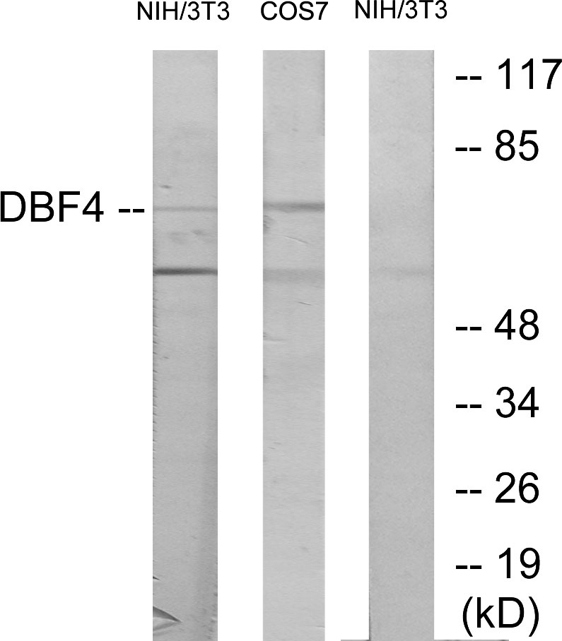
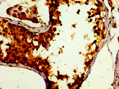
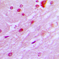
Bio-Connect
Anti-DBF4 Antibody

Anti-DBF4 Antibody

A99446
Antibodies.com
ApplicationsWestern Blot, ELISA
Product group Antibodies
Sign in to order and to see your custom pricing.
Large volume orders?
Order with a bulk request

Overview

  • Supplier
    Antibodies.com
  • Product Name
    Anti-DBF4 Antibody
  • Delivery Days Customer
    7
  • Applications
    Western Blot, ELISA
  • Certification
    Research Use Only
  • Clonality
    Polyclonal
  • Conjugate
    Unconjugated
  • Host
    Rabbit
  • Isotype
    IgG
  • Scientific Description
    Rabbit polyclonal antibody to DBF4.
  • UNSPSC
    12352203

Related products

Immunohistochemistry of paraffin-embedded human testis tissue using CSB-PA887010LA01HU at dilution of 1:100
Product group Antibodies
Cusabio
DBF4 AntibodyCSB-PA887010LA01HU
ApplicationsELISA, ImmunoHistoChemistry
TargetDBF4
  • SizePrice
Product group Antibodies
Bioss Antibodies
ApplicationsWestern Blot
TargetDBF4
  • SizePrice
Product group Antibodies
Biomatik
ApplicationsELISA, ImmunoHistoChemistry
TargetDBF4
  • SizePrice
Product group Antibodies
MedChem Express
TargetDBF4
  • SizePrice
ICC/IF analysis of formalin-fixed HepG2 cells using GTX55192 DBF4 antibody. Red : Primary antibody Blue : DAPI Permeabilization : 0.1% Triton X-100 in TBS for 5-10 minutes
Product group Antibodies
GeneTex
ApplicationsImmunoFluorescence, Western Blot, ImmunoCytoChemistry, ImmunoHistoChemistry, ImmunoHistoChemistry Paraffin
TargetDBF4
  • SizePrice
Immunofluorescent staining of human cell line RH-30 shows localization to nucleoplasm & nuclear bodies.
Product group Antibodies
Atlas Antibodies
ApplicationsImmunoCytoChemistry
TargetDBF4
  • SizePrice
Figure 1. Western blot analysis of DBF4 using anti-DBF4 antibody (A01348-2). Electrophoresis was performed on a 5-20% SDS-PAGE gel at 70V (Stacking gel) / 90V (Resolving gel) for 2-3 hours. The sample well of each lane was loaded with 30 ug of sample under reducing conditions. Lane 1: human HepG2 whole cell lysates, Lane 2: human SH-SY5Y whole cell lysates, Lane 3: rat PC-12 whole cell lysates, Lane 4: mouse NIH/3T3 whole cell lysates. After electrophoresis, proteins were transferred to a nitrocellulose membrane at 150 mA for 50-90 minutes. Blocked the membrane with 5% non-fat milk/TBS for 1.5 hour at RT. The membrane was incubated with rabbit anti-DBF4 antigen affinity purified polyclonal antibody (Catalog # A01348-2) at 0.5 microg/mL overnight at 4°C, then washed with TBS-0.1%Tween 3 times with 5 minutes each and probed with a goat anti-rabbit IgG-HRP secondary antibody at a dilution of 1:5000 for 1.5 hour at RT. The signal is developed using an Enhanced Chemiluminescent detection (ECL) kit (Catalog # EK1002) with Tanon 5200 system. A specific band was detected for DBF4 at approximately 80 kDa. The expected band size for DBF4 is at 80 kDa.
Product group Antibodies
Boster Bio
Anti-DBF4 Antibody Picoband(r)A01348-2-CARRIER-FREE
ApplicationsFlow Cytometry, ImmunoFluorescence, Western Blot, ELISA, ImmunoCytoChemistry
TargetDBF4
  • SizePrice
Product group Antibodies
RayBiotech
ApplicationsWestern Blot
TargetDBF4
  • SizePrice
Product group Antibodies
LifeSpan BioSciences
TargetDBF4
  • SizePrice