


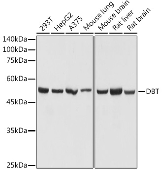
Figure 1. Western blot analysis of DBT using anti-DBT antibody (A01145-2). Electrophoresis was performed on a 5-20% SDS-PAGE gel at 70V (Stacking gel) / 90V (Resolving gel) for 2-3 hours. The sample well of each lane was loaded with 30 ug of sample under reducing conditions. Lane 1: human SiHa whole cell lysates, Lane 2: human A431 whole cell lysates, Lane 3: human Hacat whole cell lysates, Lane 4: human Jurkat whole cell lysates, Lane 5: rat stomach tissue lysates, Lane 6: rat kidney tissue lysates, Lane 7: mouse stomach tissue lysates, Lane 8: mouse kidney tissue lysates. After electrophoresis, proteins were transferred to a nitrocellulose membrane at 150 mA for 50-90 minutes. Blocked the membrane with 5% non-fat milk/TBS for 1.5 hour at RT. The membrane was incubated with rabbit anti-DBT antigen affinity purified polyclonal antibody (A01145-2) at 0.5 microg/mL overnight at 4°C, then washed with TBS-0.1%Tween 3 times with 5 minutes each and probed with a goat anti-rabbit IgG-HRP secondary antibody at a dilution of 1:5000 for 1.5 hour at RT. The signal is developed using an Enhanced Chemiluminescent detection (ECL) kit (Catalog # EK1002) with Tanon 5200 system. A specific band was detected for DBT at approximately 53 kDa. The expected band size for DBT is at 53 kDa.
 and anti-Beta Tubulin antibody (M01857-3). DBT was detected in an immunocytochemical section of U2OS cells. Enzyme antigen retrieval was performed using IHC enzyme antigen retrieval reagent (AR0022) for 15 mins. The cells were blocked with 10% goat serum. And then incubated with 5 microg/mL rabbit anti-DBT Antibody (A01145-2) and mouse anti-Beta Tubulin antibody (M01857-3) overnight at 4°C. DyLight®488 Conjugated Goat Anti-Rabbit IgG (BA1127) and DyLight®550 Conjugated Goat Anti-Mouse IgG (BA1133) were used as secondary antibody at 1:500 dilution and incubated for 30 minutes at 37°C. The section was counterstained with DAPI. Visualize using a fluorescence microscope and filter sets appropriate for the label used.")
. Lane 1: A431 whole cell lysates (30ug), Lane 2: Rabbit control IgG instead of anti-DBT antibody in A431 whole cell lysate, Lane 3: anti-DBT antibody (2microg) + A431 whole cell lysate (500microg). After electrophoresis, proteins were transferred to a membrane. Then the membrane was incubated with rabbit anti-DBT antigen affinity purified polyclonal antibody (A01145-2) at a dilution of 0.5 microg/mL and probed with a mouse anti-rabbit IgG-HRP secondary antibody (Light Chain). The signal is developed using ECL Plus Western Blotting Substrate (Catalog # AR1197). A specific band was detected for DBT at approximately 53 kDa. The expected band size for DBT is at 53 kDa.")
. Overlay histogram showing SiHa cells stained with A01145-2 (Blue line). To facilitate intracellular staining, cells were fixed with 4% paraformaldehyde and permeabilized with permeabilization buffer. The cells were blocked with 10% normal goat serum. And then incubated with rabbit anti-DBT Antibody (A01145-2, 1 microg/1x106 cells) for 30 min at 20°C. DyLight®488 conjugated goat anti-rabbit IgG (BA1127, 5-10 microg/1x106 cells) was used as secondary antibody for 30 minutes at 20°C. Isotype control antibody (Green line) was rabbit IgG (1 microg/1x106) used under the same conditions. Unlabelled sample without incubation with primary antibody and secondary antibody (Red line) was used as a blank control.")
Figure 1. Western blot analysis of DBT using anti-DBT antibody (A01145-2). Electrophoresis was performed on a 5-20% SDS-PAGE gel at 70V (Stacking gel) / 90V (Resolving gel) for 2-3 hours. The sample well of each lane was loaded with 30 ug of sample under reducing conditions. Lane 1: human SiHa whole cell lysates, Lane 2: human A431 whole cell lysates, Lane 3: human Hacat whole cell lysates, Lane 4: human Jurkat whole cell lysates, Lane 5: rat stomach tissue lysates, Lane 6: rat kidney tissue lysates, Lane 7: mouse stomach tissue lysates, Lane 8: mouse kidney tissue lysates. After electrophoresis, proteins were transferred to a nitrocellulose membrane at 150 mA for 50-90 minutes. Blocked the membrane with 5% non-fat milk/TBS for 1.5 hour at RT. The membrane was incubated with rabbit anti-DBT antigen affinity purified polyclonal antibody (A01145-2) at 0.5 microg/mL overnight at 4°C, then washed with TBS-0.1%Tween 3 times with 5 minutes each and probed with a goat anti-rabbit IgG-HRP secondary antibody at a dilution of 1:5000 for 1.5 hour at RT. The signal is developed using an Enhanced Chemiluminescent detection (ECL) kit (Catalog # EK1002) with Tanon 5200 system. A specific band was detected for DBT at approximately 53 kDa. The expected band size for DBT is at 53 kDa.
Anti-DBT Antibody Picoband(r)
A01145-2-CARRIER-FREE
ApplicationsFlow Cytometry, ImmunoFluorescence, ImmunoPrecipitation, Western Blot, ELISA, ImmunoCytoChemistry
Product group Antibodies
ReactivityHuman, Mouse, Rat
TargetDBT
Overview
- SupplierBoster Bio
- Product NameAnti-DBT Antibody Picoband(r)
- Delivery Days Customer9
- ApplicationsFlow Cytometry, ImmunoFluorescence, ImmunoPrecipitation, Western Blot, ELISA, ImmunoCytoChemistry
- CertificationResearch Use Only
- ClonalityPolyclonal
- Concentration500 ug/ml
- Gene ID1629
- Target nameDBT
- Target descriptiondihydrolipoamide branched chain transacylase E2
- Target synonymsBCATE2, BCKAD-E2, BCKADE2, BCKDH-E2, BCOADC-E2, E2, E2B, lipoamide acyltransferase component of branched-chain alpha-keto acid dehydrogenase complex, mitochondrial, 52 kDa mitochondrial autoantigen of primary biliary cirrhosis, BCKAD E2 subunit, E2 component of branched chain alpha-keto acid dehydrogenase complex, branched chain 2-oxo-acid dehydrogenase complex component E2, branched chain acyltransferase, E2 component, branched-chain alpha-keto acid dehydrogenase complex component E2, dihydrolipoamide acetyltransferase component of branched-chain alpha-keto acid dehydrogenase complex, dihydrolipoyl transacylase, dihydrolipoyllysine-residue (2-methylpropanoyl)transferase, lipoamide acyltransferase component of mitochondrial branched-chain alpha-keto acid dehydrogenase complex, mitochondrial branched chain alpha-keto acid dehydrogenase transacylase subunit (E2b)
- HostRabbit
- IsotypeIgG
- Protein IDP11182
- Protein NameLipoamide acyltransferase component of branched-chain alpha-keto acid dehydrogenase complex, mitochondrial
- Scientific DescriptionBoster Bio Anti-DBT Antibody Picoband® catalog # A01145-2. Tested in WB, ICC/IF, IP, Flow Cytometry, ELISA applications. This antibody reacts with Human, Mouse, Rat. The brand Picoband indicates this is a premium antibody that guarantees superior quality, high affinity, and strong signals with minimal background in Western blot applications. Only our best-performing antibodies are designated as Picoband, ensuring unmatched performance.
- ReactivityHuman, Mouse, Rat
- Storage Instruction-20°C,2°C to 8°C
- UNSPSC12352203
Related products
Product group Antibodies
DBT Polyclonal AntibodyCAC15648
ApplicationsWestern Blot, ELISA
ReactivityMouse
TargetDBT
- SizePrice
Product group Antibodies
Anti-DBT AntibodyA308791
ApplicationsImmunoFluorescence, Western Blot, ImmunoCytoChemistry, ImmunoHistoChemistry
ReactivityHuman, Mouse, Rat
- SizePrice
Product group Antibodies
References
DBT antibodyGTX113412
ApplicationsImmunoFluorescence, Western Blot, ImmunoCytoChemistry, ImmunoHistoChemistry, ImmunoHistoChemistry Paraffin
ReactivityHuman, Mouse, Rat
TargetDBT
- SizePrice
Product group Antibodies

DBT / E2 Antibody (HRP)LS-C318563
ApplicationsWestern Blot, ELISA
ReactivityVirus
TargetDBT
- SizePrice

Product group Antibodies
Anti-DBT AntibodyHPA026481
ApplicationsWestern Blot, ImmunoHistoChemistry
ReactivityHuman
TargetDBT
- SizePrice
Product group Antibodies
DBT AntibodyCSB-PA006530LA01HU
ApplicationsWestern Blot, ELISA
ReactivityHuman, Mouse
TargetDBT
- SizePrice