


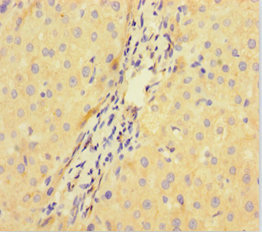
Immunohistochemical staining of human testis shows moderate cytoplasmic positivity in Leydig cells.
Immunohistochemical staining of human testis shows moderate cytoplasmic positivity in Leydig cells.
Anti-DCAF1 Antibody
HPA036142
ApplicationsImmunoHistoChemistry
Product group Antibodies
ReactivityHuman
TargetDCAF1
Overview
- SupplierAtlas Antibodies
- Product NameAnti-DCAF1 Antibody
- Delivery Days Customer4
- ApplicationsImmunoHistoChemistry
- CertificationResearch Use Only
- ClonalityPolyclonal
- ConjugateUnconjugated
- Gene ID9730
- Target nameDCAF1
- Target descriptionDDB1 and CUL4 associated factor 1
- Target synonymsRIP, VPRBP, DDB1- and CUL4-associated factor 1, HIV-1 Vpr-binding protein, Vpr (HIV-1) binding protein, Vpr-binding protein, protein VPRBP, serine/threonine-protein kinase VPRBP, vpr-interacting protein
- HostRabbit
- IsotypeIgG
- Protein IDQ9Y4B6
- Protein NameDDB1- and CUL4-associated factor 1
- Scientific DescriptionRecombinant Protein Epitope Signature Tag (PrEST) antigen sequence
- ReactivityHuman
- Storage Instruction-20°C,2°C to 8°C
- UNSPSC41116161
Datasheet
MSDS
Related products
Product group Antibodies
ApplicationsFlow Cytometry, ImmunoFluorescence, ImmunoHistoChemistry, ImmunoHistoChemistry Frozen, ImmunoHistoChemistry Paraffin
ReactivityHuman, Mouse, Rat
TargetDCAF1
- SizePrice
Product group Antibodies
DCAF1 AntibodyCSB-PA896722LA01HU
ApplicationsImmunoFluorescence, ELISA, ImmunoHistoChemistry
ReactivityHuman
TargetDCAF1
- SizePrice

Product group Antibodies
Anti-VPRBP/DCAF1 Antibody Picoband(r)A31776-1-CARRIER-FREE
ApplicationsFlow Cytometry, ImmunoFluorescence, Western Blot, ELISA, ImmunoCytoChemistry, ImmunoHistoChemistry
ReactivityHuman, Mouse, Rat
TargetDCAF1
- SizePrice
Product group Antibodies

VPRBP AntibodyLS-C679991
ApplicationsImmunoFluorescence, ELISA, ImmunoHistoChemistry, ImmunoHistoChemistry Paraffin
ReactivityHuman
TargetDCAF1
- SizePrice

Product group Antibodies
Anti-DCAF1 AntibodyHPA052445
ApplicationsImmunoCytoChemistry
ReactivityHuman
TargetDCAF1
- SizePrice

Product group Antibodies
Anti-DCAF1 AntibodyHPA053203
ApplicationsImmunoCytoChemistry, ImmunoHistoChemistry
ReactivityHuman
TargetDCAF1
- SizePrice