


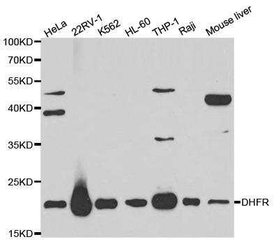
Figure 1. Western blot analysis of DHFR using anti-DHFR antibody (M00813-1). Electrophoresis was performed on a 5-20% SDS-PAGE gel at 70V (Stacking gel) / 90V (Resolving gel) for 2-3 hours. The sample well of each lane was loaded with 50ug of sample under reducing conditions. Lane 1: human K562 whole cell lysates, Lane 2: human SGC-7901 whole cell lysates, Lane 3: human Hela whole cell lysates, Lane 4: human SW620 whole cell lysates, Lane 5: rat kidney tissue lysates. After Electrophoresis, proteins were transferred to a Nitrocellulose membrane at 150mA for 50-90 minutes. Blocked the membrane with 5% Non-fat Milk/ TBS for 1.5 hour at RT. The membrane was incubated with mouse anti-DHFR antigen affinity purified monoclonal antibody (Catalog # M00813-1) at 0.5 microg/mL overnight at 4°C, then washed with TBS-0.1%Tween 3 times with 5 minutes each and probed with a goat anti-mouse IgG-HRP secondary antibody at a dilution of 1:10000 for 1.5 hour at RT. The signal is developed using an Enhanced Chemiluminescent detection (ECL) kit (Catalog # EK1001) with Tanon 5200 system. A specific band was detected for DHFR at approximately 22KD. The expected band size for DHFR is at 22KD.
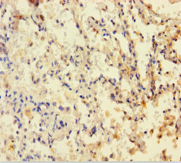
. DHFR was detected in paraffin-embedded section of human rectal cancer tissue. Heat mediated antigen retrieval was performed in EDTA buffer (pH8.0, epitope retrieval solution). The tissue section was blocked with 10% goat serum. The tissue section was then incubated with 1microg/ml mouse anti-DHFR Antibody (M00813-1) overnight at 4°C. Biotinylated goat anti-mouse IgG was used as secondary antibody and incubated for 30 minutes at 37°C. The tissue section was developed using Strepavidin-Biotin-Complex (SABC) (Catalog # SA1021) with DAB as the chromogen.")
. DHFR was detected in paraffin-embedded section of rat gaster tissue. Heat mediated antigen retrieval was performed in EDTA buffer (pH8.0, epitope retrieval solution). The tissue section was blocked with 10% goat serum. The tissue section was then incubated with 1microg/ml mouse anti-DHFR Antibody (M00813-1) overnight at 4°C. Biotinylated goat anti-mouse IgG was used as secondary antibody and incubated for 30 minutes at 37°C. The tissue section was developed using Strepavidin-Biotin-Complex (SABC) (Catalog # SA1021) with DAB as the chromogen.")
Figure 1. Western blot analysis of DHFR using anti-DHFR antibody (M00813-1). Electrophoresis was performed on a 5-20% SDS-PAGE gel at 70V (Stacking gel) / 90V (Resolving gel) for 2-3 hours. The sample well of each lane was loaded with 50ug of sample under reducing conditions. Lane 1: human K562 whole cell lysates, Lane 2: human SGC-7901 whole cell lysates, Lane 3: human Hela whole cell lysates, Lane 4: human SW620 whole cell lysates, Lane 5: rat kidney tissue lysates. After Electrophoresis, proteins were transferred to a Nitrocellulose membrane at 150mA for 50-90 minutes. Blocked the membrane with 5% Non-fat Milk/ TBS for 1.5 hour at RT. The membrane was incubated with mouse anti-DHFR antigen affinity purified monoclonal antibody (Catalog # M00813-1) at 0.5 microg/mL overnight at 4°C, then washed with TBS-0.1%Tween 3 times with 5 minutes each and probed with a goat anti-mouse IgG-HRP secondary antibody at a dilution of 1:10000 for 1.5 hour at RT. The signal is developed using an Enhanced Chemiluminescent detection (ECL) kit (Catalog # EK1001) with Tanon 5200 system. A specific band was detected for DHFR at approximately 22KD. The expected band size for DHFR is at 22KD.
Anti-DHFR Antibody Picoband(r) (monoclonal, 3C8)
M00813-1-DYLIGHT594
ApplicationsWestern Blot, ImmunoHistoChemistry
Product group Antibodies
ReactivityHuman, Rat
TargetDHFR
Overview
- SupplierBoster Bio
- Product NameAnti-DHFR Antibody Picoband(r) (monoclonal, 3C8)
- Delivery Days Customer9
- ApplicationsWestern Blot, ImmunoHistoChemistry
- CertificationResearch Use Only
- ClonalityMonoclonal
- Clone ID3C8
- Concentration500 ug/ml
- ConjugateOther Conjugate
- Gene ID1719
- Target nameDHFR
- Target descriptiondihydrofolate reductase
- Target synonymsDHFR1, DHFRP1, DYR, dihydrofolate reductase
- HostMouse
- IsotypeIgG2b
- Protein IDP00374
- Protein NameDihydrofolate reductase
- Scientific DescriptionBoster Bio Anti-DHFR Antibody Picoband® (monoclonal, 3C8) catalog # M00813-1. Tested in IHC, WB applications. This antibody reacts with Human, Rat. The brand Picoband indicates this is a premium antibody that guarantees superior quality, high affinity, and strong signals with minimal background in Western blot applications. Only our best-performing antibodies are designated as Picoband, ensuring unmatched performance.
- ReactivityHuman, Rat
- Storage Instruction-20°C,2°C to 8°C
- UNSPSC12352203
Related products
Product group Antibodies
Anti-DHFR Antibody144-01607
ApplicationsImmunoFluorescence, Western Blot
ReactivityHuman, Mouse
TargetDHFR
- SizePrice
Product group Antibodies
Dhfr Polyclonal AntibodyCAC11829
ApplicationsImmunoFluorescence, ELISA, ImmunoHistoChemistry
TargetDHFR
- SizePrice
Product group Antibodies
DHFR Recombinant Antibody, AbBy Fluor-350 ConjugatedBSM-61547R-BF350
ApplicationsImmunoFluorescence, Western Blot, ImmunoCytoChemistry
ReactivityHuman, Mouse, Rat
TargetDHFR
- SizePrice
Product group Antibodies
DHFR AntibodyCSB-PA006847HA01HU
ApplicationsImmunoFluorescence, ELISA, ImmunoHistoChemistry
ReactivityHuman
TargetDHFR
- SizePrice
Product group Antibodies
Anti-DHFR AntibodyA29840
ApplicationsWestern Blot, ImmunoHistoChemistry
ReactivityHuman, Mouse, Rat
- SizePrice
Product group Antibodies

DHFR AntibodyLS-C830690
ApplicationsELISA, ImmunoHistoChemistry
ReactivityHuman, Mouse, Rat
TargetDHFR
- SizePrice
Product group Antibodies
References
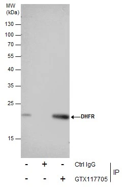
DHFR antibodyGTX117705
ApplicationsImmunoFluorescence, ImmunoPrecipitation, Western Blot, ImmunoCytoChemistry, ImmunoHistoChemistry, ImmunoHistoChemistry Paraffin
ReactivityHuman, Rat
TargetDHFR
- SizePrice

Product group Antibodies
Anti-Dihydrofolate reductase (DHFR) Antibody Picoband(r)PB9175-CARRIER-FREE
ApplicationsFlow Cytometry, ImmunoFluorescence, Western Blot, ImmunoCytoChemistry, ImmunoHistoChemistry
ReactivityHuman, Mouse, Rat
TargetDHFR
- SizePrice