


Figure 1. Western blot analysis of DLL4 using anti-DLL4 antibody (A00875-4). Electrophoresis was performed on a 5-20% SDS-PAGE gel at 70V (Stacking gel) / 90V (Resolving gel) for 2-3 hours. The sample well of each lane was loaded with 30 ug of sample under reducing conditions. Lane 1: human A431 whole cell lysates, Lane 2: human 293T whole cell lysates, Lane 3: human Caco-2 whole cell lysates, Lane 4: human PC-3 whole cell lysates, Lane 5: rat brain tissue lysates, Lane 6: mouse brain tissue lysates. After electrophoresis, proteins were transferred to a nitrocellulose membrane at 150 mA for 50-90 minutes. Blocked the membrane with 5% non-fat milk/TBS for 1.5 hour at RT. The membrane was incubated with rabbit anti-DLL4 antigen affinity purified polyclonal antibody (Catalog # A00875-4) at 0.5 microg/mL overnight at 4°C, then washed with TBS-0.1%Tween 3 times with 5 minutes each and probed with a goat anti-rabbit IgG-HRP secondary antibody at a dilution of 1:5000 for 1.5 hour at RT. The signal is developed using an Enhanced Chemiluminescent detection (ECL) kit (Catalog # EK1002) with Tanon 5200 system. A specific band was detected for DLL4 at approximately 80 kDa. The expected band size for DLL4 is at 75 kDa.
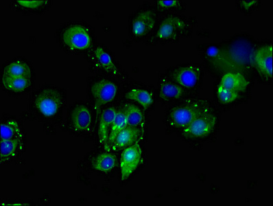
. Overlay histogram showing HepG2 cells stained with A00875-4 (Blue line). The cells were fixed with 4% paraformaldehyde and blocked with 10% normal goat serum. And then incubated with rabbit anti-DLL4 Antibody (A00875-4, 1 microg/1x106 cells) for 30 min at 20°C. DyLight®488 conjugated goat anti-rabbit IgG (BA1127, 5-10 microg/1x106 cells) was used as secondary antibody for 30 minutes at 20°C. Isotype control antibody (Green line) was rabbit IgG (1 microg/1x106) used under the same conditions. Unlabelled sample (Red line) was also used as a control.")
Figure 1. Western blot analysis of DLL4 using anti-DLL4 antibody (A00875-4). Electrophoresis was performed on a 5-20% SDS-PAGE gel at 70V (Stacking gel) / 90V (Resolving gel) for 2-3 hours. The sample well of each lane was loaded with 30 ug of sample under reducing conditions. Lane 1: human A431 whole cell lysates, Lane 2: human 293T whole cell lysates, Lane 3: human Caco-2 whole cell lysates, Lane 4: human PC-3 whole cell lysates, Lane 5: rat brain tissue lysates, Lane 6: mouse brain tissue lysates. After electrophoresis, proteins were transferred to a nitrocellulose membrane at 150 mA for 50-90 minutes. Blocked the membrane with 5% non-fat milk/TBS for 1.5 hour at RT. The membrane was incubated with rabbit anti-DLL4 antigen affinity purified polyclonal antibody (Catalog # A00875-4) at 0.5 microg/mL overnight at 4°C, then washed with TBS-0.1%Tween 3 times with 5 minutes each and probed with a goat anti-rabbit IgG-HRP secondary antibody at a dilution of 1:5000 for 1.5 hour at RT. The signal is developed using an Enhanced Chemiluminescent detection (ECL) kit (Catalog # EK1002) with Tanon 5200 system. A specific band was detected for DLL4 at approximately 80 kDa. The expected band size for DLL4 is at 75 kDa.
Anti-DLL4 Antibody Picoband(r)
A00875-4-CARRIER-FREE
ApplicationsFlow Cytometry, Western Blot, ELISA
Product group Antibodies
ReactivityHuman, Mouse, Rat
TargetDLL4
Overview
- SupplierBoster Bio
- Product NameAnti-DLL4 Antibody Picoband(r)
- Delivery Days Customer9
- ApplicationsFlow Cytometry, Western Blot, ELISA
- CertificationResearch Use Only
- ClonalityPolyclonal
- Concentration500 ug/ml
- Gene ID54567
- Target nameDLL4
- Target descriptiondelta like canonical Notch ligand 4
- Target synonymsAOS6, delta4, hdelta2, delta-like protein 4, delta 4, delta ligand 4, delta-like 4 homolog, delta-like 4 protein, drosophila Delta homolog 4, notch ligand DLL4, notch ligand delta-2
- HostRabbit
- IsotypeIgG
- Protein IDQ9NR61
- Protein NameDelta-like protein 4
- Scientific DescriptionBoster Bio Anti-DLL4 Antibody Picoband® catalog # A00875-4. Tested in ELISA, Flow Cytometry, WB applications. This antibody reacts with Human, Mouse, Rat. The brand Picoband indicates this is a premium antibody that guarantees superior quality, high affinity, and strong signals with minimal background in Western blot applications. Only our best-performing antibodies are designated as Picoband, ensuring unmatched performance.
- ReactivityHuman, Mouse, Rat
- Storage Instruction-20°C,2°C to 8°C
- UNSPSC12352203
Related products

Product group Antibodies
anti-DLL4 (human), mAb (DL86-3AG)AG-20A-0080
ApplicationsWestern Blot, ELISA
ReactivityHuman
TargetDLL4
- SizePrice
Product group Antibodies

Anti-DLL4 [61B]Ab02657-10.0
ApplicationsImmunoFluorescence, ELISA, ImmunoHistoChemistry, Neutralisation/Blocking
ReactivityHuman
TargetDLL4
- SizePrice
Product group Antibodies
Anti-DLL4 Antibody144-62568
ApplicationsImmunoFluorescence, Western Blot
ReactivityHuman, Mouse, Rat
TargetDLL4
- SizePrice
Product group Antibodies
DLL4 Polyclonal AntibodyBS-6044R
ApplicationsImmunoFluorescence, ELISA, ImmunoCytoChemistry, ImmunoHistoChemistry, ImmunoHistoChemistry Frozen, ImmunoHistoChemistry Paraffin
ReactivityBovine, Canine, Equine, Human, Mouse, Porcine, Rat
TargetDLL4
- SizePrice
Product group Antibodies
DLL4 AntibodyCSB-PA006949LA01HU
ApplicationsImmunoFluorescence, Western Blot, ELISA
ReactivityHuman
TargetDLL4
- SizePrice
Product group Antibodies

Goat anti-DLL4EB08011
ApplicationsWestern Blot, ELISA
ReactivityHuman, Mouse, Rat
TargetDLL4
- SizePrice
Product group Antibodies
DLL4 Polyclonal AntibodyCAC13569
ApplicationsImmunoFluorescence, ELISA
TargetDLL4
- SizePrice
Product group Antibodies

DLL4 AntibodyLS-C401606
ApplicationsELISA, ImmunoHistoChemistry
ReactivityHuman, Mouse
TargetDLL4
- SizePrice

Product group Antibodies
Anti-DLL4 AntibodyHPA023392
ApplicationsImmunoHistoChemistry
ReactivityHuman
TargetDLL4
- SizePrice
![Whole cell extract (30 μg) was separated by 7.5% SDS-PAGE, and the membrane was blotted with DLL4 antibody [N1N2], N-term (GTX109649) diluted at 1:2000.](https://www.genetex.com/upload/website/prouct_img/normal/GTX109649/GTX109649_39974_20151105_WB_w_23060500_574.webp)
Product group Antibodies
DLL4 antibody [N1N2], N-termGTX109649
ApplicationsWestern Blot
ReactivityHuman
TargetDLL4
- SizePrice