


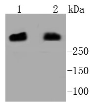
Figure 1. Western blot analysis of PRKDC using anti-PRKDC antibody (PA1970). Electrophoresis was performed on a 5-20% SDS-PAGE gel at 70V (Stacking gel) / 90V (Resolving gel) for 2-3 hours. The sample well of each lane was loaded with 30 ug of sample under reducing conditions. Lane 1: human K562 whole cell lysates, Lane 2: human HEK293 whole cell lysates. After electrophoresis, proteins were transferred to a nitrocellulose membrane at 150 mA for 50-90 minutes. Blocked the membrane with 5% non-fat milk/TBS for 1.5 hour at RT. The membrane was incubated with rabbit anti-PRKDC antigen affinity purified polyclonal antibody (Catalog # PA1970) at 0.5 microg/mL overnight at 4°C, then washed with TBS-0.1%Tween 3 times with 5 minutes each and probed with a goat anti-rabbit IgG-HRP secondary antibody at a dilution of 1:5000 for 1.5 hour at RT. The signal is developed using an Enhanced Chemiluminescent detection (ECL) kit (Catalog # EK1002) with Tanon 5200 system. A specific band was detected for PRKDC at approximately 460 kDa. The expected band size for PRKDC is at 469 kDa.
Figure 1. Western blot analysis of PRKDC using anti-PRKDC antibody (PA1970). Electrophoresis was performed on a 5-20% SDS-PAGE gel at 70V (Stacking gel) / 90V (Resolving gel) for 2-3 hours. The sample well of each lane was loaded with 30 ug of sample under reducing conditions. Lane 1: human K562 whole cell lysates, Lane 2: human HEK293 whole cell lysates. After electrophoresis, proteins were transferred to a nitrocellulose membrane at 150 mA for 50-90 minutes. Blocked the membrane with 5% non-fat milk/TBS for 1.5 hour at RT. The membrane was incubated with rabbit anti-PRKDC antigen affinity purified polyclonal antibody (Catalog # PA1970) at 0.5 microg/mL overnight at 4°C, then washed with TBS-0.1%Tween 3 times with 5 minutes each and probed with a goat anti-rabbit IgG-HRP secondary antibody at a dilution of 1:5000 for 1.5 hour at RT. The signal is developed using an Enhanced Chemiluminescent detection (ECL) kit (Catalog # EK1002) with Tanon 5200 system. A specific band was detected for PRKDC at approximately 460 kDa. The expected band size for PRKDC is at 469 kDa.
Anti-DNA PKcs Antibody
PA1970
ApplicationsWestern Blot
Product group Antibodies
ReactivityHuman
TargetPRKDC
Overview
- SupplierBoster Bio
- Product NameAnti-DNA PKcs Antibody
- Delivery Days Customer9
- Application Supplier NoteWB: The detection limit for PRKDC is approximately 2.5ng/lane under reducing conditions. Tested Species: In-house tested species with positive results. Predicted Species: Species predicted to be fit for the product based on sequence similarities. Other applications have not been tested. Optimal dilutions should be determined by end users.
- ApplicationsWestern Blot
- Applications SupplierWB
- CertificationResearch Use Only
- ClonalityPolyclonal
- Concentration500 ug/ml
- Gene ID5591
- Target namePRKDC
- Target descriptionprotein kinase, DNA-activated, catalytic subunit
- Target synonymsDNA-PKC, DNA-PKcs, DNAPK, DNAPKc, DNPK1, HYRC, HYRC1, IMD26, S473K, XRCC7, p350, DNA-dependent protein kinase catalytic subunit, DNA-PK catalytic subunit, hyper-radiosensitivity of murine scid mutation, complementing 1, p460, protein kinase, DNA-activated, catalytic polypeptide, ser-473 kinase
- HostRabbit
- IsotypeIgG
- Protein IDP78527
- Protein NameDNA-dependent protein kinase catalytic subunit
- Scientific DescriptionBoster Bio Anti-DNA PKcs/PRKDC Antibody catalog # PA1970. Tested in WB applications. This antibody reacts with Human. The brand Picoband indicates this is a premium antibody that guarantees superior quality, high affinity, and strong signals with minimal background in Western blot applications. Only our best-performing antibodies are designated as Picoband, ensuring unmatched performance.
- ReactivityHuman
- Reactivity SupplierHuman
- Storage Instruction-20°C,2°C to 8°C
- UNSPSC12352203
Datasheet
MSDS
Related products
Product group Antibodies
DNA PKcs Recombinant AntibodyBSM-52493R
ApplicationsImmunoFluorescence, Western Blot, ImmunoCytoChemistry, ImmunoHistoChemistry, ImmunoHistoChemistry Frozen, ImmunoHistoChemistry Paraffin
ReactivityHuman, Mouse, Rat
TargetPRKDC
- SizePrice
Product group Antibodies
PRKDC Polyclonal AntibodyCAC14750
ApplicationsWestern Blot, ELISA
TargetPRKDC
- SizePrice
Product group Antibodies
Anti-DNA PKcs AntibodyA12159
ApplicationsImmunoFluorescence, Western Blot, ImmunoCytoChemistry, ImmunoHistoChemistry
ReactivityHuman, Mouse
- SizePrice
Product group Antibodies
Anti-PRKDC Antibody144-07716
ApplicationsImmunoFluorescence, Western Blot, ImmunoHistoChemistry
ReactivityHuman, Mouse
TargetPRKDC
- SizePrice
Product group Antibodies
References
DNA-PKcs antibody [C3], C-termGTX109673
ApplicationsImmunoFluorescence, Western Blot, ImmunoCytoChemistry, ImmunoHistoChemistry, ImmunoHistoChemistry Paraffin
ReactivityHuman
TargetPRKDC
- SizePrice
Product group Antibodies
ApplicationsWestern Blot, ImmunoHistoChemistry
ReactivityHuman
TargetPRKDC
- SizePrice
Product group Antibodies

ApplicationsImmunoFluorescence, Western Blot, ImmunoCytoChemistry, ImmunoHistoChemistry, ImmunoHistoChemistry Paraffin
ReactivityHuman, Monkey
TargetPRKDC
- SizePrice

Product group Antibodies
Anti-PRKDC AntibodyHPA035174
ApplicationsImmunoCytoChemistry, ImmunoHistoChemistry
ReactivityHuman
TargetPRKDC
- SizePrice