


Figure 1. Western blot analysis of DTR/HBEGF using anti-DTR/HBEGF antibody (A01759-3). Electrophoresis was performed on a 5-20% SDS-PAGE gel at 70V (Stacking gel) / 90V (Resolving gel) for 2-3 hours. The sample well of each lane was loaded with 30 ug of sample under reducing conditions. Lane 1: human Hela whole cell lysates, Lane 2: human K562 whole cell lysates, Lane 3: human Jurkat whole cell lysates, Lane 4: human U251 whole cell lysates, Lane 5: rat C6 whole cell lysates, Lane 6: mouse lung tissue lysates. After electrophoresis, proteins were transferred to a nitrocellulose membrane at 150 mA for 50-90 minutes. Blocked the membrane with 5% non-fat milk/TBS for 1.5 hour at RT. The membrane was incubated with rabbit anti-DTR/HBEGF antigen affinity purified polyclonal antibody (Catalog # A01759-3) at 0.5 microg/mL overnight at 4°C, then washed with TBS-0.1%Tween 3 times with 5 minutes each and probed with a goat anti-rabbit IgG-HRP secondary antibody at a dilution of 1:5000 for 1.5 hour at RT. The signal is developed using an Enhanced Chemiluminescent detection (ECL) kit (Catalog # EK1002) with Tanon 5200 system. A specific band was detected for DTR/HBEGF at approximately 23 kDa. The expected band size for DTR/HBEGF is at 23 kDa.
. DTR/HBEGF was detected in a paraffin-embedded section of human lung adenocarcinoma tissue. Heat mediated antigen retrieval was performed in EDTA buffer (pH 8.0, epitope retrieval solution). The tissue section was blocked with 10% goat serum. The tissue section was then incubated with 2 microg/ml rabbit anti-DTR/HBEGF Antibody (A01759-3) overnight at 4°C. Peroxidase Conjugated Goat Anti-rabbit IgG was used as secondary antibody and incubated for 30 minutes at 37°C. The tissue section was developed using HRP Conjugated Rabbit IgG Super Vision Assay Kit (Catalog # SV0002) with DAB as the chromogen.")
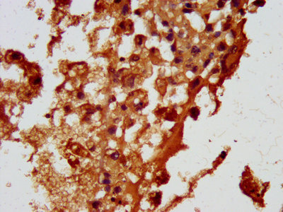
. DTR/HBEGF was detected in a paraffin-embedded section of human appendiceal adenocarcinoma tissue. Heat mediated antigen retrieval was performed in EDTA buffer (pH 8.0, epitope retrieval solution). The tissue section was blocked with 10% goat serum. The tissue section was then incubated with 2 microg/ml rabbit anti-DTR/HBEGF Antibody (A01759-3) overnight at 4°C. Peroxidase Conjugated Goat Anti-rabbit IgG was used as secondary antibody and incubated for 30 minutes at 37°C. The tissue section was developed using HRP Conjugated Rabbit IgG Super Vision Assay Kit (Catalog # SV0002) with DAB as the chromogen.")
. DTR/HBEGF was detected in a paraffin-embedded section of mouse lung tissue. Heat mediated antigen retrieval was performed in EDTA buffer (pH 8.0, epitope retrieval solution). The tissue section was blocked with 10% goat serum. The tissue section was then incubated with 2 microg/ml rabbit anti-DTR/HBEGF Antibody (A01759-3) overnight at 4°C. Peroxidase Conjugated Goat Anti-rabbit IgG was used as secondary antibody and incubated for 30 minutes at 37°C. The tissue section was developed using HRP Conjugated Rabbit IgG Super Vision Assay Kit (Catalog # SV0002) with DAB as the chromogen.")
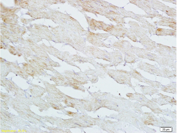
. DTR/HBEGF was detected in a paraffin-embedded section of rat respiratory smooth muscle tissue. Heat mediated antigen retrieval was performed in EDTA buffer (pH 8.0, epitope retrieval solution). The tissue section was blocked with 10% goat serum. The tissue section was then incubated with 2 microg/ml rabbit anti-DTR/HBEGF Antibody (A01759-3) overnight at 4°C. Peroxidase Conjugated Goat Anti-rabbit IgG was used as secondary antibody and incubated for 30 minutes at 37°C. The tissue section was developed using HRP Conjugated Rabbit IgG Super Vision Assay Kit (Catalog # SV0002) with DAB as the chromogen.")
. DTR/HBEGF was detected in a paraffin-embedded section of human colorectal adenocarcinoma tissue. Heat mediated antigen retrieval was performed in EDTA buffer (pH 8.0, epitope retrieval solution). The tissue section was blocked with 10% goat serum. The tissue section was then incubated with 5 microg/mL rabbit anti-DTR/HBEGF Antibody (A01759-3) overnight at 4°C. DyLight®550 Conjugated Goat Anti-Rabbit IgG (BA1135) was used as secondary antibody at 1:100 dilution and incubated for 30 minutes at 37°C. The section was counterstained with DAPI. Visualize using a fluorescence microscope and filter sets appropriate for the label used.")
. Overlay histogram showing Jurkat cells stained with A01759-3 (Blue line). The cells were fixed with 4% paraformaldehyde and blocked with 10% normal goat serum. And then incubated with rabbit anti-DTR/HBEGF Antibody (A01759-3, 1 microg/1x106 cells) for 30 min at 20°C. DyLight®488 conjugated goat anti-rabbit IgG (BA1127, 5-10 microg/1x106 cells) was used as secondary antibody for 30 minutes at 20°C. Isotype control antibody (Green line) was rabbit IgG (1 microg/1x106) used under the same conditions. Unlabelled sample without incubation with primary antibody and secondary antibody (Red line) was used as a blank control.")
Figure 1. Western blot analysis of DTR/HBEGF using anti-DTR/HBEGF antibody (A01759-3). Electrophoresis was performed on a 5-20% SDS-PAGE gel at 70V (Stacking gel) / 90V (Resolving gel) for 2-3 hours. The sample well of each lane was loaded with 30 ug of sample under reducing conditions. Lane 1: human Hela whole cell lysates, Lane 2: human K562 whole cell lysates, Lane 3: human Jurkat whole cell lysates, Lane 4: human U251 whole cell lysates, Lane 5: rat C6 whole cell lysates, Lane 6: mouse lung tissue lysates. After electrophoresis, proteins were transferred to a nitrocellulose membrane at 150 mA for 50-90 minutes. Blocked the membrane with 5% non-fat milk/TBS for 1.5 hour at RT. The membrane was incubated with rabbit anti-DTR/HBEGF antigen affinity purified polyclonal antibody (Catalog # A01759-3) at 0.5 microg/mL overnight at 4°C, then washed with TBS-0.1%Tween 3 times with 5 minutes each and probed with a goat anti-rabbit IgG-HRP secondary antibody at a dilution of 1:5000 for 1.5 hour at RT. The signal is developed using an Enhanced Chemiluminescent detection (ECL) kit (Catalog # EK1002) with Tanon 5200 system. A specific band was detected for DTR/HBEGF at approximately 23 kDa. The expected band size for DTR/HBEGF is at 23 kDa.
Anti-DTR/HBEGF Antibody Picoband(r)
A01759-3-CARRIER-FREE
ApplicationsFlow Cytometry, ImmunoFluorescence, Western Blot, ImmunoHistoChemistry
Product group Antibodies
ReactivityHuman, Mouse, Rat
TargetHBEGF
Overview
- SupplierBoster Bio
- Product NameAnti-DTR/HBEGF Antibody Picoband(r)
- Delivery Days Customer9
- ApplicationsFlow Cytometry, ImmunoFluorescence, Western Blot, ImmunoHistoChemistry
- CertificationResearch Use Only
- ClonalityPolyclonal
- Concentration500 ug/ml
- Gene ID1839
- Target nameHBEGF
- Target descriptionheparin binding EGF like growth factor
- Target synonymsDTR, DTS, DTSF, HEGFL, proheparin-binding EGF-like growth factor, diphtheria toxin receptor (heparin-binding EGF-like growth factor), diphtheria toxin receptor (heparin-binding epidermal growth factor-like growth factor), heparin-binding epidermal growth factor
- HostRabbit
- IsotypeIgG
- Protein IDQ99075
- Protein NameProheparin-binding EGF-like growth factor
- Scientific DescriptionBoster Bio Anti-DTR/HBEGF Antibody Picoband® catalog # A01759-3. Tested in Flow Cytometry, IF, IHC, WB applications. This antibody reacts with Human, Mouse, Rat. The brand Picoband indicates this is a premium antibody that guarantees superior quality, high affinity, and strong signals with minimal background in Western blot applications. Only our best-performing antibodies are designated as Picoband, ensuring unmatched performance.
- ReactivityHuman, Mouse, Rat
- Storage Instruction-20°C,2°C to 8°C
- UNSPSC12352203
Related products
Product group Antibodies
Anti-HBEGF Antibody144-01695
ApplicationsWestern Blot
ReactivityHuman, Mouse, Rat
TargetHBEGF
- SizePrice
Product group Antibodies

Anti-HB-EGF [KM3566]AB04154-1.1
ApplicationsFlow Cytometry, Western Blot, ELISA, Neutralisation/Blocking
ReactivityHuman
TargetHBEGF
- SizePrice
Product group Antibodies
References
HBEGF Polyclonal AntibodyBS-3576R
ApplicationsImmunoFluorescence, ELISA, ImmunoCytoChemistry, ImmunoHistoChemistry, ImmunoHistoChemistry Paraffin
ReactivityBovine, Canine, Chicken, Equine, Human, Mouse, Porcine, Rabbit, Rat
TargetHBEGF
- SizePrice
Product group Antibodies
HBEGF AntibodyCSB-PA857429LA01HU
ApplicationsImmunoFluorescence, ELISA, ImmunoHistoChemistry
ReactivityHuman
TargetHBEGF
- SizePrice
Product group Antibodies
Hbegf Polyclonal AntibodyCAC11350
ApplicationsImmunoFluorescence, ELISA, ImmunoHistoChemistry
TargetHBEGF
- SizePrice
Product group Antibodies

HBEGF / HB EGF AntibodyLS-C408338
ApplicationsWestern Blot, ImmunoHistoChemistry
ReactivityHuman, Mouse
TargetHBEGF
- SizePrice
![ICC/IF analysis of various samples using GTX00702 HB EGF antibody [4G10]. Vero-H : Vero cells overexpressing human HB-EGF protein Vero-mH : Vero cells overexpressing mouse HB-EGF protein Fixation : 4% PFA](https://www.genetex.com/upload/website/prouct_img/normal/GTX00702/GTX00702_20191104_ICC-IF_w_23053121_434.webp)
Product group Antibodies
HB EGF antibody [4G10]GTX00702
ApplicationsImmunoFluorescence, ImmunoPrecipitation, Western Blot, ImmunoCytoChemistry, ImmunoHistoChemistry, Neutralisation/Blocking
ReactivityHuman
TargetHBEGF
- SizePrice

Product group Antibodies
Anti-HBEGF AntibodyHPA053243
ApplicationsImmunoHistoChemistry
ReactivityHuman
TargetHBEGF
- SizePrice