


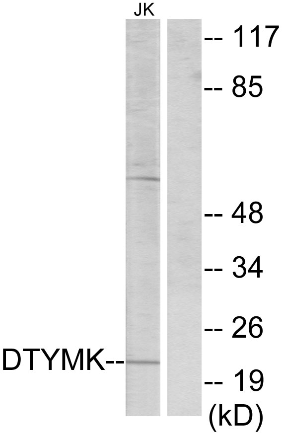
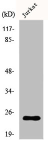
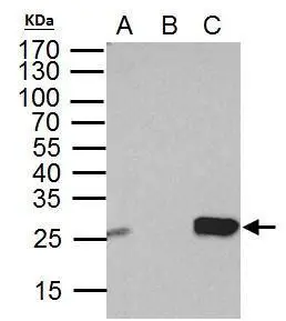
Figure 1. Western blot analysis of DTYMK using anti-DTYMK antibody (M12209-1). Electrophoresis was performed on a 5-20% SDS-PAGE gel at 70V (Stacking gel) / 90V (Resolving gel) for 2-3 hours. The sample well of each lane was loaded with 30 ug of sample under reducing conditions. Lane 1: human A549 whole cell lysates, Lane 2: human Hela whole cell lysates, Lane 3: human PC-3 whole cell lysates, Lane 4: human U20S whole cell lysates. After electrophoresis, proteins were transferred to a nitrocellulose membrane at 150 mA for 50-90 minutes. Blocked the membrane with 5% non-fat milk/TBS for 1.5 hour at RT. The membrane was incubated with rabbit anti-DTYMK antigen affinity purified monoclonal antibody (Catalog # M12209-1) at 1:500 overnight at 4°C, then washed with TBS-0.1%Tween 3 times with 5 minutes each and probed with a goat anti-rabbit IgG-HRP secondary antibody at a dilution of 1:500 for 1.5 hour at RT. The signal is developed using an Enhanced Chemiluminescent detection (ECL) kit (Catalog # EK1002) with Tanon 5200 system. A specific band was detected for DTYMK at approximately 24 kDa. The expected band size for DTYMK is at 24 kDa.
Figure 1. Western blot analysis of DTYMK using anti-DTYMK antibody (M12209-1). Electrophoresis was performed on a 5-20% SDS-PAGE gel at 70V (Stacking gel) / 90V (Resolving gel) for 2-3 hours. The sample well of each lane was loaded with 30 ug of sample under reducing conditions. Lane 1: human A549 whole cell lysates, Lane 2: human Hela whole cell lysates, Lane 3: human PC-3 whole cell lysates, Lane 4: human U20S whole cell lysates. After electrophoresis, proteins were transferred to a nitrocellulose membrane at 150 mA for 50-90 minutes. Blocked the membrane with 5% non-fat milk/TBS for 1.5 hour at RT. The membrane was incubated with rabbit anti-DTYMK antigen affinity purified monoclonal antibody (Catalog # M12209-1) at 1:500 overnight at 4°C, then washed with TBS-0.1%Tween 3 times with 5 minutes each and probed with a goat anti-rabbit IgG-HRP secondary antibody at a dilution of 1:500 for 1.5 hour at RT. The signal is developed using an Enhanced Chemiluminescent detection (ECL) kit (Catalog # EK1002) with Tanon 5200 system. A specific band was detected for DTYMK at approximately 24 kDa. The expected band size for DTYMK is at 24 kDa.
Anti-DTYMK Rabbit Monoclonal Antibody
M12209-1
ApplicationsFlow Cytometry, ImmunoFluorescence, Western Blot, ImmunoCytoChemistry, ImmunoHistoChemistry
Product group Antibodies
ReactivityHuman
TargetDTYMK
Overview
- SupplierBoster Bio
- Product NameAnti-DTYMK Rabbit Monoclonal Antibody
- Delivery Days Customer9
- ApplicationsFlow Cytometry, ImmunoFluorescence, Western Blot, ImmunoCytoChemistry, ImmunoHistoChemistry
- CertificationResearch Use Only
- ClonalityMonoclonal
- Clone ID29D22
- Gene ID1841
- Target nameDTYMK
- Target descriptiondeoxythymidylate kinase
- Target synonymsCDC8, CONPM, PP3731, TMPK, TYMK, thymidylate kinase, dTMP kinase, deoxythymidylate kinase (thymidylate kinase)
- HostRabbit
- IsotypeIgG
- Protein IDP23919
- Protein NameThymidylate kinase
- Scientific DescriptionBoster Bio Anti-DTYMK Rabbit Monoclonal Antibody catalog # M12209-1. Tested in WB, IHC, ICC/IF, Flow Cytometry applications. This antibody reacts with Human.
- ReactivityHuman
- Storage Instruction-20°C
- UNSPSC12352203
Related products
Product group Antibodies
Anti-DTYMK AntibodyA99653
ApplicationsWestern Blot, ELISA, ImmunoHistoChemistry
ReactivityHuman
- SizePrice
Product group Antibodies
Anti-DTYMK Antibody144-06370
ApplicationsImmunoFluorescence, Western Blot
ReactivityHuman, Mouse, Rat
TargetDTYMK
- SizePrice
Product group Antibodies

Thymidylate Kinase AntibodyLS-C830578
ApplicationsWestern Blot, ELISA
ReactivityHuman, Mouse
TargetDTYMK
- SizePrice
Product group Antibodies
ApplicationsFlow Cytometry, Western Blot, ImmunoCytoChemistry
ReactivityHuman
TargetDTYMK
- SizePrice
Product group Antibodies
DTYMK AntibodyCSB-PA002214
ApplicationsWestern Blot, ELISA, ImmunoHistoChemistry
ReactivityHuman
TargetDTYMK
- SizePrice
Product group Antibodies
ApplicationsImmunoPrecipitation, Western Blot, ImmunoCytoChemistry, ImmunoHistoChemistry
ReactivityPorcine
TargetDTYMK
- SizePrice

Product group Antibodies
Anti-DTYMK AntibodyHPA042593
ApplicationsWestern Blot, ImmunoHistoChemistry
ReactivityHuman
TargetDTYMK
- SizePrice
Product group Antibodies
DTYMK antibodyGTX114371
ApplicationsImmunoPrecipitation, Western Blot
ReactivityHuman
TargetDTYMK
- SizePrice
Product group Antibodies
Anti-DTYMK AntibodyCAB6370
ApplicationsImmunoFluorescence, Western Blot, ELISA, ImmunoCytoChemistry
ReactivityHuman
TargetDTYMK
- SizePrice