


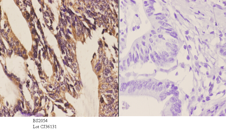
Immunohistochemical staining of human colon shows cytoplasmic positivity in glandular cells.

Immunohistochemical staining of human colon shows cytoplasmic positivity in glandular cells.
Anti-DYRK1A Antibody
HPA015810
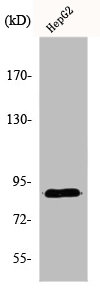
ApplicationsWestern Blot, ImmunoHistoChemistry
Product group Antibodies
ReactivityHuman
TargetDYRK1A
Overview
- SupplierAtlas Antibodies
- Product NameAnti-DYRK1A Antibody
- Delivery Days Customer4
- ApplicationsWestern Blot, ImmunoHistoChemistry
- CertificationResearch Use Only
- ClonalityPolyclonal
- ConjugateUnconjugated
- Gene ID1859
- Target nameDYRK1A
- Target descriptiondual specificity tyrosine phosphorylation regulated kinase 1A
- Target synonymsDYRK, DYRK1, HP86, MNB, MNBH, MRD7, dual specificity tyrosine-phosphorylation-regulated kinase 1A, MNB/DYRK protein kinase, dual specificity YAK1-related kinase, dual specificity tyrosine-(Y)-phosphorylation regulated kinase 1A, mnb protein kinase homolog hp86, protein kinase minibrain homolog, serine/threonine kinase MNB, serine/threonine-specific protein kinase
- HostRabbit
- IsotypeIgG
- Protein IDQ13627
- Protein NameDual specificity tyrosine-phosphorylation-regulated kinase 1A
- Scientific DescriptionRecombinant Protein Epitope Signature Tag (PrEST) antigen sequence
- ReactivityHuman
- Storage Instruction-20°C,2°C to 8°C
- UNSPSC41116161
Datasheet
MSDS
Related products
Product group Antibodies
ApplicationsImmunoFluorescence, Western Blot, ImmunoHistoChemistry
ReactivityHuman, Mouse, Rat
- SizePrice

Product group Antibodies
Anti-DYRK1A Antibody Picoband(r)A00878-1-CARRIER-FREE
ApplicationsImmunoFluorescence, Western Blot, ELISA, ImmunoCytoChemistry
ReactivityHuman, Rat
TargetDYRK1A
- SizePrice
Product group Antibodies
Anti-DYRK1A Antibody144-00595
ApplicationsWestern Blot
ReactivityHuman, Mouse, Rat
TargetDYRK1A
- SizePrice
Product group Antibodies
DYRK1A AntibodyCSB-PA002232
ApplicationsImmunoFluorescence, Western Blot, ELISA, ImmunoHistoChemistry
ReactivityHuman, Mouse, Rat
TargetDYRK1A
- SizePrice
Product group Antibodies
References

Goat anti-DYRK1AEB11483
ApplicationsImmunoPrecipitation, Western Blot, ELISA
ReactivityBovine, Canine, Human, Mouse, Porcine, Rat
TargetDYRK1A
- SizePrice
Product group Antibodies
ApplicationsImmunoPrecipitation, Western Blot, ImmunoCytoChemistry, ImmunoHistoChemistry
ReactivityMouse, Porcine, Rat
TargetDYRK1A
- SizePrice
Product group Antibodies

DYRK / DYRK1A AntibodyLS-C408229
ApplicationsWestern Blot
ReactivityHuman, Mouse, Rat
TargetDYRK1A
- SizePrice