


Figure 1. Western blot analysis of Dysferlin using anti-Dysferlin antibody (M01234). Electrophoresis was performed on a 5-20% SDS-PAGE gel at 70V (Stacking gel) / 90V (Resolving gel) for 2-3 hours. The sample well of each lane was loaded with 30 ug of sample under reducing conditions. Lane 1: mouse skeletal muscle tissue lysates. After electrophoresis, proteins were transferred to a nitrocellulose membrane at 150 mA for 50-90 minutes. Blocked the membrane with 5% non-fat milk/TBS for 1.5 hour at RT. The membrane was incubated with rabbit anti-Dysferlin antigen affinity purified monoclonal antibody (Catalog # M01234) at 1:500 overnight at 4°C, then washed with TBS-0.1%Tween 3 times with 5 minutes each and probed with a goat anti-rabbit IgG-HRP secondary antibody at a dilution of 1:5000 for 1.5 hour at RT. The signal is developed using an Enhanced Chemiluminescent detection (ECL) kit (Catalog # EK1002) with Tanon 5200 system. A specific band was detected for Dysferlin at approximately 280 kDa. The expected band size for Dysferlin is at 237 kDa.
Figure 1. Western blot analysis of Dysferlin using anti-Dysferlin antibody (M01234). Electrophoresis was performed on a 5-20% SDS-PAGE gel at 70V (Stacking gel) / 90V (Resolving gel) for 2-3 hours. The sample well of each lane was loaded with 30 ug of sample under reducing conditions. Lane 1: mouse skeletal muscle tissue lysates. After electrophoresis, proteins were transferred to a nitrocellulose membrane at 150 mA for 50-90 minutes. Blocked the membrane with 5% non-fat milk/TBS for 1.5 hour at RT. The membrane was incubated with rabbit anti-Dysferlin antigen affinity purified monoclonal antibody (Catalog # M01234) at 1:500 overnight at 4°C, then washed with TBS-0.1%Tween 3 times with 5 minutes each and probed with a goat anti-rabbit IgG-HRP secondary antibody at a dilution of 1:5000 for 1.5 hour at RT. The signal is developed using an Enhanced Chemiluminescent detection (ECL) kit (Catalog # EK1002) with Tanon 5200 system. A specific band was detected for Dysferlin at approximately 280 kDa. The expected band size for Dysferlin is at 237 kDa.
Anti-Dysferlin Rabbit Monoclonal Antibody
M01234
ApplicationsImmunoFluorescence, Western Blot, ImmunoCytoChemistry, ImmunoHistoChemistry
Product group Antibodies
ReactivityHuman, Mouse
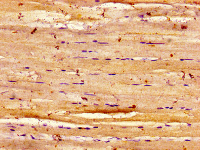
TargetDYSF
Overview
- SupplierBoster Bio
- Product NameAnti-Dysferlin Rabbit Monoclonal Antibody
- Delivery Days Customer9
- ApplicationsImmunoFluorescence, Western Blot, ImmunoCytoChemistry, ImmunoHistoChemistry
- CertificationResearch Use Only
- ClonalityMonoclonal
- Clone IDEGO-4
- Gene ID8291
- Target nameDYSF
- Target descriptiondysferlin
- Target synonymsFER1L1, LGMD2B, LGMDR2, MMD1, dysferlin, dystrophy-associated fer-1-like 1, fer-1-like family member 1, fer-1-like protein 1, limb girdle muscular dystrophy 2B (autosomal recessive)
- HostRabbit
- IsotypeIgG
- Protein IDO75923
- Protein NameDysferlin
- Scientific DescriptionBoster Bio Anti-Dysferlin Rabbit Monoclonal Antibody catalog # M01234. Tested in WB, IHC, ICC/IF applications. This antibody reacts with Human, Mouse.
- ReactivityHuman, Mouse
- Storage Instruction-20°C
- UNSPSC12352203
Datasheet
MSDS
Related products

Product group Antibodies
Anti-Dysferlin AntibodyA97816
ApplicationsWestern Blot, ELISA
ReactivityHuman, Mouse
- SizePrice


Product group Antibodies
Anti-Dysferlin/DYSF Antibody Picoband(r)A01234-3-CARRIER-FREE
ApplicationsFlow Cytometry, ImmunoFluorescence, Western Blot, ELISA, ImmunoCytoChemistry, ImmunoHistoChemistry
ReactivityHuman, Mouse, Rat
TargetDYSF
- SizePrice

Product group Antibodies
Anti-DYSF AntibodyAMAB91667
ApplicationsWestern Blot, ImmunoHistoChemistry
ReactivityHuman
TargetDYSF
- SizePrice
Product group Antibodies

Dysferlin Antibody (aa105-278)LS-C673588
ApplicationsELISA, ImmunoHistoChemistry, ImmunoHistoChemistry Paraffin
ReactivityHuman
TargetDYSF
- SizePrice
Product group Antibodies
Dysferlin Recombinant AntibodyBSM-61236R
ApplicationsImmunoFluorescence, Western Blot, ImmunoCytoChemistry, ImmunoHistoChemistry, ImmunoHistoChemistry Frozen, ImmunoHistoChemistry Paraffin
TargetDYSF
- SizePrice
Product group Antibodies
DYSF AntibodyCSB-PA007314LA01HU
ApplicationsImmunoFluorescence, ELISA, ImmunoHistoChemistry
ReactivityHuman
TargetDYSF
- SizePrice
Product group Antibodies

Goat anti-dysferlinEB12749
ApplicationsELISA
ReactivityBovine, Human, Porcine, Rat
TargetDYSF
- SizePrice
Product group Antibodies
DYSF Polyclonal AntibodyCAC13026
ApplicationsImmunoFluorescence, ELISA, ImmunoHistoChemistry
TargetDYSF
- SizePrice