


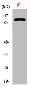
Figure 1. Western blot analysis of E Cadherin using anti-E Cadherin antibody (PB9561). Electrophoresis was performed on a 5-20% SDS-PAGE gel at 70V (Stacking gel) / 90V (Resolving gel) for 2-3 hours. The sample well of each lane was loaded with 30 ug of sample under reducing conditions. Lane 1: human MCF-7 whole cell lysates, Lane 2: human A431 whole cell lysates. After electrophoresis, proteins were transferred to a nitrocellulose membrane at 150 mA for 50-90 minutes. Blocked the membrane with 5% non-fat milk/TBS for 1.5 hour at RT. The membrane was incubated with rabbit anti-E Cadherin antigen affinity purified polyclonal antibody (Catalog # PB9561) at 0.5 microg/mL overnight at 4°C, then washed with TBS-0.1%Tween 3 times with 5 minutes each and probed with a goat anti-rabbit IgG-HRP secondary antibody at a dilution of 1:5000 for 1.5 hour at RT. The signal is developed using an Enhanced Chemiluminescent detection (ECL) kit (Catalog # EK1002) with Tanon 5200 system. A specific band was detected for E Cadherin at approximately 130 kDa. The expected band size for E Cadherin is at 97 kDa.
. E Cadherin was detected in paraffin-embedded section of Human Placenta Tissue. Heat mediated antigen retrieval was performed in citrate buffer (pH6, epitope retrieval solution) for 20 mins. The tissue section was blocked with 10% goat serum. The tissue section was then incubated with 1microg/ml rabbit anti-E Cadherin Antibody (PB9561) overnight at 4°C. Biotinylated goat anti-rabbit IgG was used as secondary antibody and incubated for 30 minutes at 37°C. The tissue section was developed using Strepavidin-Biotin-Complex (SABC)(Catalog # SA1022) with DAB as the chromogen.")
. E Cadherin was detected in frozen section of Human Placenta Tissue. The tissue section was blocked with 10% goat serum. The tissue section was then incubated with 1microg/ml rabbit anti-E Cadherin Antibody (PB9561) overnight at 4°C. Biotinylated goat anti-rabbit IgG was used as secondary antibody and incubated for 30 minutes at 37°C. The tissue section was developed using Strepavidin-Biotin-Complex (SABC)(Catalog # SA1022) with DAB as the chromogen.")
. E Cadherin was detected in paraffin-embedded section of mouse intestine tissues. Heat mediated antigen retrieval was performed in citrate buffer (pH6, epitope retrieval solution) for 20 mins. The tissue section was blocked with 10% goat serum. The tissue section was then incubated with 1microg/ml rabbit anti-E Cadherin Antibody (PB9561) overnight at 4°C. Biotinylated goat anti-rabbit IgG was used as secondary antibody and incubated for 30 minutes at 37°C. The tissue section was developed using Strepavidin-Biotin-Complex (SABC)(Catalog # SA1022) with DAB as the chromogen.")
. E Cadherin was detected in paraffin-embedded section of rat intestine tissues. Heat mediated antigen retrieval was performed in citrate buffer (pH6, epitope retrieval solution) for 20 mins. The tissue section was blocked with 10% goat serum. The tissue section was then incubated with 1microg/ml rabbit anti-E Cadherin Antibody (PB9561) overnight at 4°C. Biotinylated goat anti-rabbit IgG was used as secondary antibody and incubated for 30 minutes at 37°C. The tissue section was developed using Strepavidin-Biotin-Complex (SABC)(Catalog # SA1022) with DAB as the chromogen.")
. E Cadherin was detected in frozen section of mouse intestine tissues. The tissue section was blocked with 10% goat serum. The tissue section was then incubated with 1microg/ml rabbit anti-E Cadherin Antibody (PB9561) overnight at 4°C. Biotinylated goat anti-rabbit IgG was used as secondary antibody and incubated for 30 minutes at 37°C. The tissue section was developed using Strepavidin-Biotin-Complex (SABC)(Catalog # SA1022) with DAB as the chromogen.")
. E Cadherin was detected in frozen section of rat intestine tissues. The tissue section was blocked with 10% goat serum. The tissue section was then incubated with 1microg/ml rabbit anti-E Cadherin Antibody (PB9561) overnight at 4°C. Biotinylated goat anti-rabbit IgG was used as secondary antibody and incubated for 30 minutes at 37°C. The tissue section was developed using Strepavidin-Biotin-Complex (SABC)(Catalog # SA1022) with DAB as the chromogen.")
. E Cadherin was detected in frozen section of human placenta tissues. The tissue section was blocked with 10% goat serum. The tissue section was then incubated with 1microg/ml rabbit anti-E Cadherin Antibody (PB9561) overnight at 4°C. Biotinylated goat anti-rabbit IgG was used as secondary antibody and incubated for 30 minutes at 37°C. The tissue section was developed using Strepavidin-Biotin-Complex (SABC)(Catalog # SA1022) with DAB as the chromogen.")
. E Cadherin was detected in paraffin-embedded section of human ileum tissue. Heat mediated antigen retrieval was performed in citrate buffer (pH6, epitope retrieval solution) for 20 mins. The tissue section was blocked with 10% goat serum. The tissue section was then incubated with 5microg/mL rabbit anti-E Cadherin Antibody (PB9561) overnight at 4°C. Cy3 Conjugated Goat Anti-Rabbit IgG (BA1032) was used as secondary antibody at 1:100 dilution and incubated for 30 minutes at 37°C. The section was counterstained with DAPI. Visualize using a fluorescence microscope and filter sets appropriate for the label used.")
. E Cadherin was detected in paraffin-embedded section of human colon organoid tissue. Heat mediated antigen retrieval was performed in citrate buffer (pH6, epitope retrieval solution) for 20 mins. The tissue section was blocked with 10% goat serum. The tissue section was then incubated with 5microg/mL rabbit anti-E Cadherin Antibody (PB9561) overnight at 4°C. Cy3 Conjugated Goat Anti-Rabbit IgG (BA1032) was used as secondary antibody at 1:100 dilution and incubated for 30 minutes at 37°C. The section was counterstained with DAPI. Visualize using a fluorescence microscope and filter sets appropriate for the label used.")
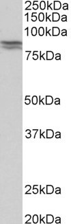
Figure 1. Western blot analysis of E Cadherin using anti-E Cadherin antibody (PB9561). Electrophoresis was performed on a 5-20% SDS-PAGE gel at 70V (Stacking gel) / 90V (Resolving gel) for 2-3 hours. The sample well of each lane was loaded with 30 ug of sample under reducing conditions. Lane 1: human MCF-7 whole cell lysates, Lane 2: human A431 whole cell lysates. After electrophoresis, proteins were transferred to a nitrocellulose membrane at 150 mA for 50-90 minutes. Blocked the membrane with 5% non-fat milk/TBS for 1.5 hour at RT. The membrane was incubated with rabbit anti-E Cadherin antigen affinity purified polyclonal antibody (Catalog # PB9561) at 0.5 microg/mL overnight at 4°C, then washed with TBS-0.1%Tween 3 times with 5 minutes each and probed with a goat anti-rabbit IgG-HRP secondary antibody at a dilution of 1:5000 for 1.5 hour at RT. The signal is developed using an Enhanced Chemiluminescent detection (ECL) kit (Catalog # EK1002) with Tanon 5200 system. A specific band was detected for E Cadherin at approximately 130 kDa. The expected band size for E Cadherin is at 97 kDa.
Anti-E Cadherin 1/CDH1 Antibody Picoband(r)
PB9561-CARRIER-FREE
ApplicationsImmunoFluorescence, Western Blot, ELISA, ImmunoCytoChemistry, ImmunoHistoChemistry, ImmunoHistoChemistry Frozen
Product group Antibodies
ReactivityHuman, Mouse, Rat
TargetCDH1
Overview
- SupplierBoster Bio
- Product NameAnti-E Cadherin 1/CDH1 Antibody Picoband(r)
- Delivery Days Customer9
- Application Supplier NoteTested Species: In-house tested species with positive results. By Heat: Boiling the paraffin sections in 10mM citrate buffer, pH6.0, for 20mins is required for the staining of formalin/paraffin sections. Other applications have not been tested. Optimal dilutions should be determined by end users.
- ApplicationsImmunoFluorescence, Western Blot, ELISA, ImmunoCytoChemistry, ImmunoHistoChemistry, ImmunoHistoChemistry Frozen
- CertificationResearch Use Only
- ClonalityPolyclonal
- Concentration500 ug/ml
- Gene ID999
- Target nameCDH1
- Target descriptioncadherin 1
- Target synonymsArc-1, BCDS1, CD324, CDHE, ECAD, LCAM, UVO, cadherin-1, CAM 120/80, E-cadherin 1, cadherin 1, E-cadherin (epithelial), cadherin 1, type 1, E-cadherin (epithelial), calcium-dependent adhesion protein, epithelial, cell-CAM 120/80, epididymis secretory sperm binding protein, epithelial cadherin, uvomorulin
- HostRabbit
- IsotypeIgG
- Protein IDP12830
- Protein NameCadherin-1
- Scientific DescriptionBoster Bio Anti-E Cadherin 1/CDH1 Antibody Picoband® catalog # PB9561. Tested in ELISA, IF, IHC, IHC-F, ICC, WB applications. This antibody reacts with Human, Mouse, Rat. The brand Picoband indicates this is a premium antibody that guarantees superior quality, high affinity, and strong signals with minimal background in Western blot applications. Only our best-performing antibodies are designated as Picoband, ensuring unmatched performance.
- ReactivityHuman, Mouse, Rat
- Storage Instruction-20°C,2°C to 8°C
- UNSPSC12352203
Related products
Product group Antibodies
CDH1 AntibodyCSB-PA002243
ApplicationsImmunoFluorescence, Western Blot, ELISA, ImmunoHistoChemistry
ReactivityHuman
TargetCDH1
- SizePrice
Product group Antibodies
Anti-E Cadherin AntibodyA83532
ApplicationsWestern Blot, ELISA, ImmunoHistoChemistry
ReactivityHuman, Mouse, Porcine, Rat
- SizePrice

Product group Antibodies
Anti-CDH1 AntibodyAMAB90862
ApplicationsWestern Blot, ImmunoCytoChemistry, ImmunoHistoChemistry
ReactivityHuman
TargetCDH1
- SizePrice
Product group Antibodies

Anti-E Cadherin [4A2]AB01700-1.1-BT
ApplicationsFlow Cytometry, ImmunoFluorescence, ImmunoPrecipitation, Western Blot, ImmunoHistoChemistry
ReactivityHuman, Mouse, Rat
TargetCDH1
- SizePrice
Product group Antibodies

CDH1 / E Cadherin Antibody (aa155-709)LS-C293089
ApplicationsImmunoPrecipitation, Western Blot, ImmunoCytoChemistry
ReactivityRat
TargetCDH1
- SizePrice
Product group Antibodies

Goat anti-CDH1 (aa662-675)EB11206
ApplicationsWestern Blot, ELISA, ImmunoHistoChemistry
ReactivityCanine, Human, Mouse, Porcine, Rat
TargetCDH1
- SizePrice
Product group Antibodies

ApplicationsFlow Cytometry, Western Blot, ImmunoCytoChemistry, ImmunoHistoChemistry, ImmunoHistoChemistry Frozen
ReactivityHuman
TargetCDH1
- SizePrice
Product group Antibodies
Cdh1 Polyclonal AntibodyCAC11436
ApplicationsImmunoFluorescence, ImmunoPrecipitation, Western Blot, ELISA, ImmunoHistoChemistry
TargetCDH1
- SizePrice