

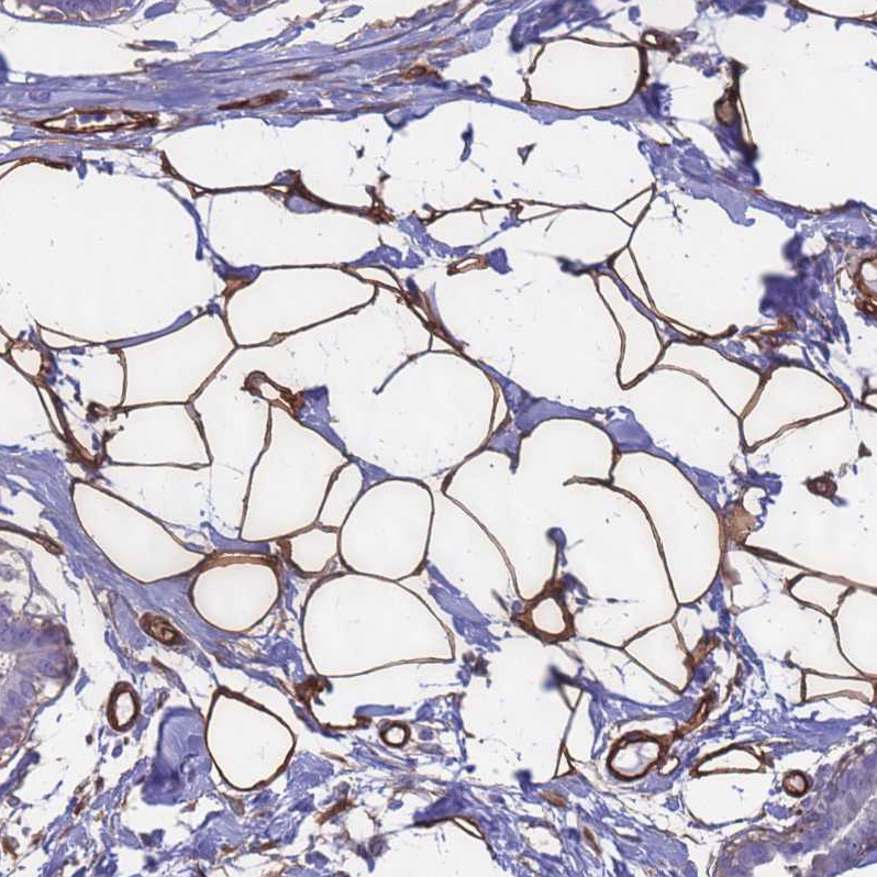
Immunohistochemical staining of human Breast shows strong membranous positivity in adipocytes.
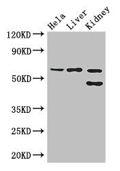
![Lane 1: Marker [kDa] 250, 130, 95, 72, 55, 36, 28, 17, 10. Lane 2: Human cell line RT-4. Lane 3: Human cell line U-251MG sp](https://atlasantibodies.s3.amazonaws.com/images/wb/hpa047394-wb-1.jpg "Lane 1: Marker [kDa] 250, 130, 95, 72, 55, 36, 28, 17, 10. Lane 2: Human cell line RT-4. Lane 3: Human cell line U-251MG sp")
Immunohistochemical staining of human Breast shows strong membranous positivity in adipocytes.
Anti-EHD2 Antibody
HPA047394
ApplicationsWestern Blot, ImmunoCytoChemistry, ImmunoHistoChemistry
Product group Antibodies
ReactivityHuman
TargetEHD2
Overview
- SupplierAtlas Antibodies
- Product NameAnti-EHD2 Antibody
- Delivery Days Customer4
- ApplicationsWestern Blot, ImmunoCytoChemistry, ImmunoHistoChemistry
- CertificationResearch Use Only
- ClonalityPolyclonal
- ConjugateUnconjugated
- Gene ID30846
- Target nameEHD2
- Target descriptionEH domain containing 2
- Target synonymsPAST2, EH domain-containing protein 2, PAST homolog 2
- HostRabbit
- IsotypeIgG
- Protein IDQ9NZN4
- Protein NameEH domain-containing protein 2
- Scientific DescriptionRecombinant Protein Epitope Signature Tag (PrEST) antigen sequence
- ReactivityHuman
- Storage Instruction-20°C,2°C to 8°C
- UNSPSC41116161
Datasheet
MSDS
Related products

Product group Antibodies
Anti-EHD2 AntibodyA90734
ApplicationsWestern Blot
ReactivityHuman, Mouse, Rat
- SizePrice
Product group Antibodies

EHD2 AntibodyLS-C748018
ApplicationsWestern Blot
ReactivityHuman, Mouse, Rat
TargetEHD2
- SizePrice
Product group Antibodies

Goat anti-EHD2EB06493
ApplicationsFlow Cytometry, ImmunoFluorescence, Western Blot, ELISA
ReactivityBovine, Canine, Human, Mouse, Rat
TargetEHD2
- SizePrice

Product group Antibodies
Anti-EHD2 Antibody Picoband(r)A04265-2-CARRIER-FREE
ApplicationsImmunoFluorescence, Western Blot, ImmunoCytoChemistry
ReactivityHuman
TargetEHD2
- SizePrice
Product group Antibodies
EHD2 AntibodyCSB-PA873710LA01HU
ApplicationsImmunoFluorescence, Western Blot, ELISA, ImmunoHistoChemistry
ReactivityHuman, Mouse
TargetEHD2
- SizePrice
Product group Antibodies
Ehd2 Polyclonal AntibodyCAC07805
ApplicationsImmunoFluorescence, Western Blot, ELISA, ImmunoHistoChemistry
ReactivityMouse
TargetEHD2
- SizePrice
Product group Antibodies
EHD2 antibodyGTX66142
ApplicationsWestern Blot
ReactivityHuman, Mouse, Rat
TargetEHD2
- SizePrice
Product group Antibodies
Anti-EHD2 Antibody144-12945
ApplicationsWestern Blot
ReactivityHuman, Mouse, Rat
TargetEHD2
- SizePrice