


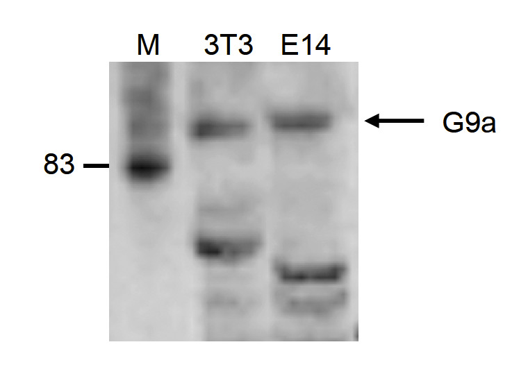
Figure 1. Western blot analysis of EHMT2/G9A using anti-EHMT2/G9A antibody (A01055-2). Electrophoresis was performed on a 5-20% SDS-PAGE gel at 70V (Stacking gel) / 90V (Resolving gel) for 2-3 hours. The sample well of each lane was loaded with 30 ug of sample under reducing conditions. Lane 1: human PC-3 whole cell lysates, Lane 2: human HEK293 whole cell lysates, Lane 3: human U87 whole cell lysates, Lane 4: human A549 whole cell lysates, Lane 5: human U20S whole cell lysates, Lane 6: rat testis tissue lysates. After electrophoresis, proteins were transferred to a nitrocellulose membrane at 150 mA for 50-90 minutes. Blocked the membrane with 5% non-fat milk/TBS for 1.5 hour at RT. The membrane was incubated with rabbit anti-EHMT2/G9A antigen affinity purified polyclonal antibody (Catalog # A01055-2) at 0.5 microg/mL overnight at 4°C, then washed with TBS-0.1%Tween 3 times with 5 minutes each and probed with a goat anti-rabbit IgG-HRP secondary antibody at a dilution of 1:5000 for 1.5 hour at RT. The signal is developed using an Enhanced Chemiluminescent detection (ECL) kit (Catalog # EK1002) with Tanon 5200 system. A specific band was detected for EHMT2/G9A at approximately 160-180 kDa. The expected band size for EHMT2/G9A is at 160-180 kDa.
. EHMT2/G9A was detected in a paraffin-embedded section of human liver cancer tissue. Heat mediated antigen retrieval was performed in EDTA buffer (pH 8.0, epitope retrieval solution). The tissue section was blocked with 10% goat serum. The tissue section was then incubated with 2 microg/ml rabbit anti-EHMT2/G9A Antibody (A01055-2) overnight at 4°C. Biotinylated goat anti-rabbit IgG was used as secondary antibody and incubated for 30 minutes at 37°C. The tissue section was developed using Strepavidin-Biotin-Complex (SABC) (Catalog # SA1022) with DAB as the chromogen.")
. EHMT2/G9A was detected in a paraffin-embedded section of human rectal cancer tissue. Heat mediated antigen retrieval was performed in EDTA buffer (pH 8.0, epitope retrieval solution). The tissue section was blocked with 10% goat serum. The tissue section was then incubated with 2 microg/ml rabbit anti-EHMT2/G9A Antibody (A01055-2) overnight at 4°C. Biotinylated goat anti-rabbit IgG was used as secondary antibody and incubated for 30 minutes at 37°C. The tissue section was developed using Strepavidin-Biotin-Complex (SABC) (Catalog # SA1022) with DAB as the chromogen.")
. EHMT2/G9A was detected in a paraffin-embedded section of mouse ovary tissue. Heat mediated antigen retrieval was performed in EDTA buffer (pH 8.0, epitope retrieval solution). The tissue section was blocked with 10% goat serum. The tissue section was then incubated with 2 microg/ml rabbit anti-EHMT2/G9A Antibody (A01055-2) overnight at 4°C. Biotinylated goat anti-rabbit IgG was used as secondary antibody and incubated for 30 minutes at 37°C. The tissue section was developed using Strepavidin-Biotin-Complex (SABC) (Catalog # SA1022) with DAB as the chromogen.")
. EHMT2/G9A was detected in an immunocytochemical section of A549 cells. Enzyme antigen retrieval was performed using IHC enzyme antigen retrieval reagent (AR0022) for 15 mins. The cells were blocked with 10% goat serum. And then incubated with 5 microg/mL rabbit anti-EHMT2/G9A Antibody (A01055-2) overnight at 4°C. DyLight®488 Conjugated Goat Anti-Rabbit IgG (BA1127) was used as secondary antibody at 1:100 dilution and incubated for 30 minutes at 37°C. The section was counterstained with DAPI. Visualize using a fluorescence microscope and filter sets appropriate for the label used.")
. Overlay histogram showing HL-60 cells stained with A01055-2 (Blue line). To facilitate intracellular staining, cells were fixed with 4% paraformaldehyde and permeabilized with permeabilization buffer. The cells were blocked with 10% normal goat serum. And then incubated with rabbit anti-EHMT2/G9A Antibody (A01055-2, 1 microg/1x106 cells) for 30 min at 20°C. DyLight®488 conjugated goat anti-rabbit IgG (BA1127, 5-10 microg/1x106 cells) was used as secondary antibody for 30 minutes at 20°C. Isotype control antibody (Green line) was rabbit IgG (1 microg/1x106) used under the same conditions. Unlabelled sample without incubation with primary antibody and secondary antibody (Red line) was used as a blank control.")
Figure 1. Western blot analysis of EHMT2/G9A using anti-EHMT2/G9A antibody (A01055-2). Electrophoresis was performed on a 5-20% SDS-PAGE gel at 70V (Stacking gel) / 90V (Resolving gel) for 2-3 hours. The sample well of each lane was loaded with 30 ug of sample under reducing conditions. Lane 1: human PC-3 whole cell lysates, Lane 2: human HEK293 whole cell lysates, Lane 3: human U87 whole cell lysates, Lane 4: human A549 whole cell lysates, Lane 5: human U20S whole cell lysates, Lane 6: rat testis tissue lysates. After electrophoresis, proteins were transferred to a nitrocellulose membrane at 150 mA for 50-90 minutes. Blocked the membrane with 5% non-fat milk/TBS for 1.5 hour at RT. The membrane was incubated with rabbit anti-EHMT2/G9A antigen affinity purified polyclonal antibody (Catalog # A01055-2) at 0.5 microg/mL overnight at 4°C, then washed with TBS-0.1%Tween 3 times with 5 minutes each and probed with a goat anti-rabbit IgG-HRP secondary antibody at a dilution of 1:5000 for 1.5 hour at RT. The signal is developed using an Enhanced Chemiluminescent detection (ECL) kit (Catalog # EK1002) with Tanon 5200 system. A specific band was detected for EHMT2/G9A at approximately 160-180 kDa. The expected band size for EHMT2/G9A is at 160-180 kDa.
Anti-EHMT2/G9A Antibody Picoband(r)
A01055-2-CARRIER-FREE
ApplicationsFlow Cytometry, ImmunoFluorescence, Western Blot, ELISA, ImmunoCytoChemistry, ImmunoHistoChemistry
Product group Antibodies
ReactivityHuman, Mouse, Rat
TargetEHMT2
Overview
- SupplierBoster Bio
- Product NameAnti-EHMT2/G9A Antibody Picoband(r)
- Delivery Days Customer9
- ApplicationsFlow Cytometry, ImmunoFluorescence, Western Blot, ELISA, ImmunoCytoChemistry, ImmunoHistoChemistry
- CertificationResearch Use Only
- ClonalityPolyclonal
- Concentration500 ug/ml
- Gene ID10919
- Target nameEHMT2
- Target descriptioneuchromatic histone lysine methyltransferase 2
- Target synonymsBAT8, C6orf30, G9A, GAT8, KMT1C, NG36, histone-lysine N-methyltransferase EHMT2, G9A histone methyltransferase, H3-K9-HMTase 3, HLA-B associated transcript 8, ankyrin repeat-containing protein, euchromatic histone-lysine N-methyltransferase 2, histone H3-K9 methyltransferase 3, histone-lysine N-methyltransferase, H3 lysine-9 specific 3, lysine N-methyltransferase 1C
- HostRabbit
- IsotypeIgG
- Protein IDQ96KQ7
- Protein NameHistone-lysine N-methyltransferase EHMT2
- Scientific DescriptionBoster Bio Anti-EHMT2/G9A Antibody Picoband® catalog # A01055-2. Tested in ELISA, Flow Cytometry, IF, IHC, ICC, WB applications. This antibody reacts with Human, Mouse, Rat. The brand Picoband indicates this is a premium antibody that guarantees superior quality, high affinity, and strong signals with minimal background in Western blot applications. Only our best-performing antibodies are designated as Picoband, ensuring unmatched performance.
- ReactivityHuman, Mouse, Rat
- Storage Instruction-20°C,2°C to 8°C
- UNSPSC12352203
Related products

Product group Antibodies
Anti-EHMT2 AntibodyA29536
ApplicationsWestern Blot, ImmunoHistoChemistry
ReactivityHuman, Mouse, Rat
- SizePrice
Product group Antibodies
Anti-EHMT2 Antibody144-01247
ApplicationsWestern Blot, ImmunoHistoChemistry
ReactivityHuman, Mouse, Rat
TargetEHMT2
- SizePrice
Product group Antibodies
ApplicationsWestern Blot, ELISA
ReactivityMouse
TargetEHMT2
- SizePrice
Product group Antibodies
EHMT2 Recombinant Antibody, Biotin ConjugatedBSM-61806R-BIOTIN
ApplicationsImmunoPrecipitation, Western Blot, ImmunoHistoChemistry, ImmunoHistoChemistry Frozen, ImmunoHistoChemistry Paraffin
ReactivityHuman, Mouse, Rat
TargetEHMT2
- SizePrice
Product group Antibodies
EHMT2 AntibodyCSB-PA007497GA01HU
ApplicationsWestern Blot, ELISA
ReactivityHuman, Mouse, Rat
TargetEHMT2
- SizePrice
Product group Antibodies

EHMT2 / G9A AntibodyLS-C331386
ApplicationsWestern Blot, ImmunoHistoChemistry
ReactivityHuman, Mouse, Rat
TargetEHMT2
- SizePrice

Product group Antibodies
Anti-EHMT2 AntibodyHPA060259
ApplicationsWestern Blot, ChIP Chromatin ImmunoPrecipitation, ImmunoCytoChemistry
ReactivityHuman
TargetEHMT2
- SizePrice
Product group Antibodies
G9a antibodyGTX128164
ApplicationsImmunoFluorescence, ImmunoPrecipitation, Western Blot, ImmunoCytoChemistry, ImmunoHistoChemistry, ImmunoHistoChemistry Paraffin
ReactivityHuman, Mouse
TargetEHMT2
- SizePrice
Product group Antibodies
Anti-MYO10 AntibodyCAB12471
ApplicationsImmunoFluorescence, Western Blot, ELISA, ImmunoCytoChemistry
ReactivityHuman
TargetEHMT2
- SizePrice