


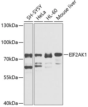
Figure 1. Western blot analysis of EIF2AK1 using anti-EIF2AK1 antibody (A05465-2). Electrophoresis was performed on a 5-20% SDS-PAGE gel at 70V (Stacking gel) / 90V (Resolving gel) for 2-3 hours. The sample well of each lane was loaded with 30 ug of sample under reducing conditions. Lane 1: human Hela whole cell lysates, Lane 2: human HepG2 whole cell lysates, Lane 3: human 293T whole cell lysates, Lane 4: rat liver tissue lysates, Lane 5: mouse liver tissue lysates. After electrophoresis, proteins were transferred to a nitrocellulose membrane at 150 mA for 50-90 minutes. Blocked the membrane with 5% non-fat milk/TBS for 1.5 hour at RT. The membrane was incubated with rabbit anti-EIF2AK1 antigen affinity purified polyclonal antibody (Catalog # A05465-2) at 0.5 microg/mL overnight at 4°C, then washed with TBS-0.1%Tween 3 times with 5 minutes each and probed with a goat anti-rabbit IgG-HRP secondary antibody at a dilution of 1:5000 for 1.5 hour at RT. The signal is developed using an Enhanced Chemiluminescent detection (ECL) kit (Catalog # EK1002) with Tanon 5200 system. A specific band was detected for EIF2AK1 at approximately 71 kDa. The expected band size for EIF2AK1 is at 71 kDa.
. EIF2AK1 was detected in a paraffin-embedded section of human glioblastoma tissue. Heat mediated antigen retrieval was performed in EDTA buffer (pH 8.0, epitope retrieval solution). The tissue section was blocked with 10% goat serum. The tissue section was then incubated with 2 microg/ml rabbit anti-EIF2AK1 Antibody (A05465-2) overnight at 4°C. Peroxidase Conjugated Goat Anti-rabbit IgG was used as secondary antibody and incubated for 30 minutes at 37°C. The tissue section was developed using HRP Conjugated Rabbit IgG Super Vision Assay Kit (Catalog # SV0002) with DAB as the chromogen.")
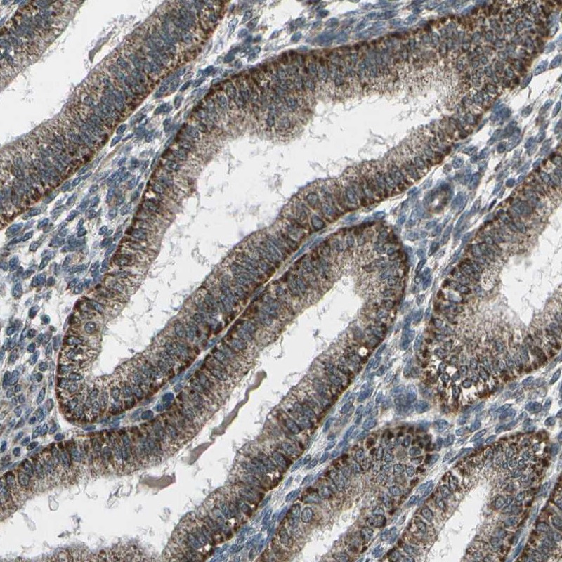
. EIF2AK1 was detected in a paraffin-embedded section of human liver cancer tissue. Heat mediated antigen retrieval was performed in EDTA buffer (pH 8.0, epitope retrieval solution). The tissue section was blocked with 10% goat serum. The tissue section was then incubated with 2 microg/ml rabbit anti-EIF2AK1 Antibody (A05465-2) overnight at 4°C. Peroxidase Conjugated Goat Anti-rabbit IgG was used as secondary antibody and incubated for 30 minutes at 37°C. The tissue section was developed using HRP Conjugated Rabbit IgG Super Vision Assay Kit (Catalog # SV0002) with DAB as the chromogen.")
. EIF2AK1 was detected in a paraffin-embedded section of human testicular seminoma tissue. Heat mediated antigen retrieval was performed in EDTA buffer (pH 8.0, epitope retrieval solution). The tissue section was blocked with 10% goat serum. The tissue section was then incubated with 2 microg/ml rabbit anti-EIF2AK1 Antibody (A05465-2) overnight at 4°C. Peroxidase Conjugated Goat Anti-rabbit IgG was used as secondary antibody and incubated for 30 minutes at 37°C. The tissue section was developed using HRP Conjugated Rabbit IgG Super Vision Assay Kit (Catalog # SV0002) with DAB as the chromogen.")
. EIF2AK1 was detected in an immunocytochemical section of U87 cells. Enzyme antigen retrieval was performed using IHC enzyme antigen retrieval reagent (AR0022) for 15 mins. The cells were blocked with 10% goat serum. And then incubated with 5 microg/mL rabbit anti-EIF2AK1 Antibody (A05465-2) overnight at 4°C. Cy3 Conjugated Goat Anti-Rabbit IgG (BA1032) was used as secondary antibody at 1:500 dilution and incubated for 30 minutes at 37°C. The section was counterstained with DAPI. Visualize using a fluorescence microscope and filter sets appropriate for the label used.")
Figure 1. Western blot analysis of EIF2AK1 using anti-EIF2AK1 antibody (A05465-2). Electrophoresis was performed on a 5-20% SDS-PAGE gel at 70V (Stacking gel) / 90V (Resolving gel) for 2-3 hours. The sample well of each lane was loaded with 30 ug of sample under reducing conditions. Lane 1: human Hela whole cell lysates, Lane 2: human HepG2 whole cell lysates, Lane 3: human 293T whole cell lysates, Lane 4: rat liver tissue lysates, Lane 5: mouse liver tissue lysates. After electrophoresis, proteins were transferred to a nitrocellulose membrane at 150 mA for 50-90 minutes. Blocked the membrane with 5% non-fat milk/TBS for 1.5 hour at RT. The membrane was incubated with rabbit anti-EIF2AK1 antigen affinity purified polyclonal antibody (Catalog # A05465-2) at 0.5 microg/mL overnight at 4°C, then washed with TBS-0.1%Tween 3 times with 5 minutes each and probed with a goat anti-rabbit IgG-HRP secondary antibody at a dilution of 1:5000 for 1.5 hour at RT. The signal is developed using an Enhanced Chemiluminescent detection (ECL) kit (Catalog # EK1002) with Tanon 5200 system. A specific band was detected for EIF2AK1 at approximately 71 kDa. The expected band size for EIF2AK1 is at 71 kDa.
Anti-EIF2AK1 Antibody Picoband(r)
A05465-2-CARRIER-FREE
ApplicationsImmunoFluorescence, Western Blot, ELISA, ImmunoCytoChemistry, ImmunoHistoChemistry
Product group Antibodies
ReactivityHuman, Mouse, Rat
TargetEIF2AK1
Overview
- SupplierBoster Bio
- Product NameAnti-EIF2AK1 Antibody Picoband(r)
- Delivery Days Customer9
- ApplicationsImmunoFluorescence, Western Blot, ELISA, ImmunoCytoChemistry, ImmunoHistoChemistry
- CertificationResearch Use Only
- ClonalityPolyclonal
- Concentration500 ug/ml
- Gene ID27102
- Target nameEIF2AK1
- Target descriptioneukaryotic translation initiation factor 2 alpha kinase 1
- Target synonymsHCR, HRI, LEMSPAD, hHRI, eukaryotic translation initiation factor 2-alpha kinase 1, heme regulated initiation factor 2 alpha kinase, heme sensitive initiation factor 2a kinase, heme-controlled repressor, heme-regulated eukaryotic initiation factor eIF-2-alpha kinase, heme-regulated inhibitor, heme-regulated repressor, hemin-controlled repressor, hemin-sensitive initiation factor 2-alpha kinase
- HostRabbit
- IsotypeIgG
- Protein IDQ9BQI3
- Protein NameEukaryotic translation initiation factor 2-alpha kinase 1
- Scientific DescriptionBoster Bio Anti-EIF2AK1 Antibody Picoband® catalog # A05465-2. Tested in ELISA, Flow Cytometry, IF, IHC, ICC, WB applications. This antibody reacts with Human, Mouse, Rat. The brand Picoband indicates this is a premium antibody that guarantees superior quality, high affinity, and strong signals with minimal background in Western blot applications. Only our best-performing antibodies are designated as Picoband, ensuring unmatched performance.
- ReactivityHuman, Mouse, Rat
- Storage Instruction-20°C,2°C to 8°C
- UNSPSC12352203
Related products
Product group Antibodies
Anti-EIF2AK1 AntibodyA98608
ApplicationsWestern Blot, ELISA
ReactivityHuman, Mouse
- SizePrice
Product group Antibodies
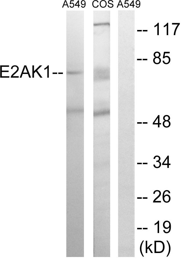
Anti-EIF2AK1 Antibody144-63086
ApplicationsWestern Blot
ReactivityHuman, Rat
TargetEIF2AK1
- SizePrice
Product group Antibodies

EIF2AK1 AntibodyLS-C831589
ApplicationsImmunoHistoChemistry
ReactivityHuman, Mouse, Rat
TargetEIF2AK1
- SizePrice

Product group Antibodies
EIF2AK1 AntibodyCSB-PA002969
ApplicationsWestern Blot, ELISA, ImmunoHistoChemistry
ReactivityHuman, Monkey, Mouse
TargetEIF2AK1
- SizePrice
Product group Antibodies
Eif2Ak1 Polyclonal AntibodyCAC07983
ApplicationsImmunoFluorescence, Western Blot, ELISA, ImmunoHistoChemistry
ReactivityRat
TargetEIF2AK1
- SizePrice
Product group Antibodies
EIF2AK1 antibodyGTX00664
ApplicationsWestern Blot
ReactivityHuman, Mouse
TargetEIF2AK1
- SizePrice
Product group Antibodies
Anti-EIF2AK1 AntibodyHPA016496
ApplicationsWestern Blot, ImmunoCytoChemistry, ImmunoHistoChemistry
ReactivityHuman
TargetEIF2AK1
- SizePrice
Product group Antibodies
Anti-EIF2AK1 AntibodyCAB3415
ApplicationsWestern Blot, ELISA
ReactivityHuman
TargetEIF2AK1
- SizePrice