


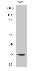
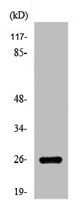
Figure 1. Western blot analysis of EIF4E using anti-EIF4E antibody (M00135). Electrophoresis was performed on a 5-20% SDS-PAGE gel at 70V (Stacking gel) / 90V (Resolving gel) for 2-3 hours. The sample well of each lane was loaded with 30 ug of sample under reducing conditions. Lane 1: human 293T whole cell lysates, Lane 2: human Hela whole cell lysates, Lane 3: human HepG2 whole cell lysates, Lane 4: human A549 whole cell lysates, Lane 5: rat RH35 whole cell lysates, Lane 6: rat PC-12 whole cell lysates, Lane 7: mouse HEPA1-2 whole cell lysates, Lane 8: mouse NIH/3T3 whole cell lysates. After electrophoresis, proteins were transferred to a nitrocellulose membrane at 150 mA for 50-90 minutes. Blocked the membrane with 5% non-fat milk/TBS for 1.5 hour at RT. The membrane was incubated with rabbit anti-EIF4E antigen affinity purified monoclonal antibody (Catalog # M00135) at 1:100 overnight at 4°C, then washed with TBS-0.1%Tween 3 times with 5 minutes each and probed with a goat anti-rabbit IgG-HRP secondary antibody at a dilution of 1:5000 for 1.5 hour at RT. The signal is developed using an Enhanced Chemiluminescent detection (ECL) kit (Catalog # EK1002) with Tanon 5200 system. A specific band was detected for EIF4E at approximately 25 kDa. The expected band size for EIF4E is at 25 kDa.
Figure 1. Western blot analysis of EIF4E using anti-EIF4E antibody (M00135). Electrophoresis was performed on a 5-20% SDS-PAGE gel at 70V (Stacking gel) / 90V (Resolving gel) for 2-3 hours. The sample well of each lane was loaded with 30 ug of sample under reducing conditions. Lane 1: human 293T whole cell lysates, Lane 2: human Hela whole cell lysates, Lane 3: human HepG2 whole cell lysates, Lane 4: human A549 whole cell lysates, Lane 5: rat RH35 whole cell lysates, Lane 6: rat PC-12 whole cell lysates, Lane 7: mouse HEPA1-2 whole cell lysates, Lane 8: mouse NIH/3T3 whole cell lysates. After electrophoresis, proteins were transferred to a nitrocellulose membrane at 150 mA for 50-90 minutes. Blocked the membrane with 5% non-fat milk/TBS for 1.5 hour at RT. The membrane was incubated with rabbit anti-EIF4E antigen affinity purified monoclonal antibody (Catalog # M00135) at 1:100 overnight at 4°C, then washed with TBS-0.1%Tween 3 times with 5 minutes each and probed with a goat anti-rabbit IgG-HRP secondary antibody at a dilution of 1:5000 for 1.5 hour at RT. The signal is developed using an Enhanced Chemiluminescent detection (ECL) kit (Catalog # EK1002) with Tanon 5200 system. A specific band was detected for EIF4E at approximately 25 kDa. The expected band size for EIF4E is at 25 kDa.
Anti-EIF4E Monoclonal Antibody
M00135
ApplicationsFlow Cytometry, ImmunoPrecipitation, Western Blot, ImmunoHistoChemistry
Product group Antibodies
ReactivityHuman, Mouse, Rat
TargetEIF4E
Overview
- SupplierBoster Bio
- Product NameAnti-EIF4E Monoclonal Antibody
- Delivery Days Customer9
- ApplicationsFlow Cytometry, ImmunoPrecipitation, Western Blot, ImmunoHistoChemistry
- CertificationResearch Use Only
- ClonalityMonoclonal
- Clone IDAFHI-5
- Gene ID1977
- Target nameEIF4E
- Target descriptioneukaryotic translation initiation factor 4E
- Target synonymsAUTS19, CBP, EIF4E1, EIF4EL1, EIF4F, eIF-4E, eukaryotic translation initiation factor 4E, eIF-4F 25 kDa subunit, eukaryotic translation initiation factor 4E-like 1, mRNA cap-binding protein
- HostRabbit
- IsotypeIgG
- Protein IDP06730
- Protein NameEukaryotic translation initiation factor 4E
- Scientific DescriptionBoster Bio Anti-EIF4E Monoclonal Antibody catalog # M00135. Tested in WB, IHC, IP, Flow Cytometry applications. This antibody reacts with Human, Mouse, Rat.
- ReactivityHuman, Mouse, Rat
- Storage Instruction-20°C
- UNSPSC12352203
Datasheet
MSDS
Related products
Product group Antibodies
Anti-eIF4E AntibodyA35900
ApplicationsWestern Blot, ELISA, ImmunoHistoChemistry
ReactivityHuman, Monkey, Mouse, Rat
- SizePrice
Product group Antibodies
Anti-EIF4E Antibody144-60024
ApplicationsImmunoFluorescence, Western Blot
ReactivityHuman
TargetEIF4E
- SizePrice

Product group Antibodies

EIF4E AntibodyLS-C746741
ApplicationsImmunoFluorescence, Western Blot
ReactivityHuman
TargetEIF4E
- SizePrice

Product group Antibodies
ApplicationsImmunoFluorescence, Western Blot, ImmunoCytoChemistry, ImmunoHistoChemistry, ImmunoHistoChemistry Frozen, ImmunoHistoChemistry Paraffin
ReactivityHuman, Mouse, Rat
TargetEIF4E
- SizePrice
Product group Antibodies
EIF4E AntibodyCSB-PA002308
ApplicationsWestern Blot, ELISA, ImmunoHistoChemistry
ReactivityHuman, Monkey, Mouse, Rat
TargetEIF4E
- SizePrice
Product group Antibodies
ApplicationsWestern Blot, ELISA, ImmunoHistoChemistry
TargetEIF4E
- SizePrice
Product group Antibodies
eIF4E antibodyGTX132092
ApplicationsImmunoFluorescence, Western Blot, ImmunoCytoChemistry, ImmunoHistoChemistry, ImmunoHistoChemistry Paraffin
ReactivityHuman, Mouse
TargetEIF4E
- SizePrice

Product group Antibodies
Anti-EIF4E AntibodyHPA051311
ApplicationsWestern Blot, ImmunoCytoChemistry
ReactivityHuman
TargetEIF4E
- SizePrice