


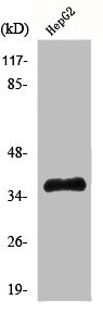
Figure 1. Western blot analysis of ELAVL2 HuB using anti-ELAVL2 HuB antibody (A06194-2). Electrophoresis was performed on a 5-20% SDS-PAGE gel at 70V (Stacking gel) / 90V (Resolving gel) for 2-3 hours. The sample well of each lane was loaded with 50ug of sample under reducing conditions. Lane 1: human T-47D whole cell lysate, Lane 2: human A549 whole cell lysate, Lane 3: human Caco-2 whole cell lysate. After Electrophoresis, proteins were transferred to a Nitrocellulose membrane at 150mA for 50-90 minutes. Blocked the membrane with 5% Non-fat Milk/ TBS for 1.5 hour at RT. The membrane was incubated with rabbit anti-ELAVL2 HuB antigen affinity purified polyclonal antibody (Catalog # A06194-2) at 0.5 microg/mL overnight at 4°C, then washed with TBS-0.1%Tween 3 times with 5 minutes each and probed with a goat anti-rabbit IgG-HRP secondary antibody at a dilution of 1:10000 for 1.5 hour at RT. The signal is developed using an Enhanced Chemiluminescent detection (ECL) kit (Catalog # EK1002) with Tanon 5200 system. A specific band was detected for ELAVL2 HuB at approximately 45KD. The expected band size for ELAVL2 HuB is at 40KD.
. ELAVL2 HuB was detected in paraffin-embedded section of mouse brain tissue. Heat mediated antigen retrieval was performed in citrate buffer (pH6, epitope retrieval solution) for 20 mins. The tissue section was blocked with 10% goat serum. The tissue section was then incubated with 1microg/ml rabbit anti-ELAVL2 HuB Antibody (A06194-2) overnight at 4°C. Biotinylated goat anti-rabbit IgG was used as secondary antibody and incubated for 30 minutes at 37°C. The tissue section was developed using Strepavidin-Biotin-Complex (SABC)(Catalog # SA1022) with DAB as the chromogen.")
. ELAVL2 HuB was detected in paraffin-embedded section of mouse brain tissue. Heat mediated antigen retrieval was performed in citrate buffer (pH6, epitope retrieval solution) for 20 mins. The tissue section was blocked with 10% goat serum. The tissue section was then incubated with 1microg/ml rabbit anti-ELAVL2 HuB Antibody (A06194-2) overnight at 4°C. Biotinylated goat anti-rabbit IgG was used as secondary antibody and incubated for 30 minutes at 37°C. The tissue section was developed using Strepavidin-Biotin-Complex (SABC)(Catalog # SA1022) with DAB as the chromogen.")
. ELAVL2 HuB was detected in paraffin-embedded section of mouse brain tissue. Heat mediated antigen retrieval was performed in citrate buffer (pH6, epitope retrieval solution) for 20 mins. The tissue section was blocked with 10% goat serum. The tissue section was then incubated with 1microg/ml rabbit anti-ELAVL2 HuB Antibody (A06194-2) overnight at 4°C. Biotinylated goat anti-rabbit IgG was used as secondary antibody and incubated for 30 minutes at 37°C. The tissue section was developed using Strepavidin-Biotin-Complex (SABC)(Catalog # SA1022) with DAB as the chromogen.")
. ELAVL2 HuB was detected in paraffin-embedded section of human glioma tissue. Heat mediated antigen retrieval was performed in citrate buffer (pH6, epitope retrieval solution) for 20 mins. The tissue section was blocked with 10% goat serum. The tissue section was then incubated with 1microg/ml rabbit anti-ELAVL2 HuB Antibody (A06194-2) overnight at 4°C. Biotinylated goat anti-rabbit IgG was used as secondary antibody and incubated for 30 minutes at 37°C. The tissue section was developed using Strepavidin-Biotin-Complex (SABC)(Catalog # SA1022) with DAB as the chromogen.")
Figure 1. Western blot analysis of ELAVL2 HuB using anti-ELAVL2 HuB antibody (A06194-2). Electrophoresis was performed on a 5-20% SDS-PAGE gel at 70V (Stacking gel) / 90V (Resolving gel) for 2-3 hours. The sample well of each lane was loaded with 50ug of sample under reducing conditions. Lane 1: human T-47D whole cell lysate, Lane 2: human A549 whole cell lysate, Lane 3: human Caco-2 whole cell lysate. After Electrophoresis, proteins were transferred to a Nitrocellulose membrane at 150mA for 50-90 minutes. Blocked the membrane with 5% Non-fat Milk/ TBS for 1.5 hour at RT. The membrane was incubated with rabbit anti-ELAVL2 HuB antigen affinity purified polyclonal antibody (Catalog # A06194-2) at 0.5 microg/mL overnight at 4°C, then washed with TBS-0.1%Tween 3 times with 5 minutes each and probed with a goat anti-rabbit IgG-HRP secondary antibody at a dilution of 1:10000 for 1.5 hour at RT. The signal is developed using an Enhanced Chemiluminescent detection (ECL) kit (Catalog # EK1002) with Tanon 5200 system. A specific band was detected for ELAVL2 HuB at approximately 45KD. The expected band size for ELAVL2 HuB is at 40KD.
Anti-ELAVL2 HuB Antibody Picoband(r)
A06194-2-CARRIER-FREE
ApplicationsWestern Blot, ImmunoHistoChemistry
Product group Antibodies
ReactivityHuman, Mouse, Rat
TargetELAVL2
Overview
- SupplierBoster Bio
- Product NameAnti-ELAVL2 HuB Antibody Picoband(r)
- Delivery Days Customer9
- ApplicationsWestern Blot, ImmunoHistoChemistry
- CertificationResearch Use Only
- ClonalityPolyclonal
- Concentration500 ug/ml
- Gene ID1993
- Target nameELAVL2
- Target descriptionELAV like RNA binding protein 2
- Target synonymsHEL-N1, HELN1, HUB, ELAV-like protein 2, ELAV (embryonic lethal, abnormal vision, Drosophila)-like 2 (Hu antigen B), ELAV like neuron-specific RNA binding protein 2, hu-antigen B, nervous system-specific RNA-binding protein Hel-N1
- HostRabbit
- IsotypeIgG
- Protein IDQ12926
- Protein NameELAV-like protein 2
- Scientific DescriptionBoster Bio Anti-ELAVL2 HuB Antibody Picoband® catalog # A06194-2. Tested in IHC, WB applications. This antibody reacts with Human, Mouse, Rat. The brand Picoband indicates this is a premium antibody that guarantees superior quality, high affinity, and strong signals with minimal background in Western blot applications. Only our best-performing antibodies are designated as Picoband, ensuring unmatched performance.
- ReactivityHuman, Mouse, Rat
- Storage Instruction-20°C,2°C to 8°C
- UNSPSC12352203
Related products
Product group Antibodies
Anti-ELAVL2 AntibodyA96904
ApplicationsWestern Blot, ELISA
ReactivityHuman, Mouse, Rat
- SizePrice
Product group Antibodies
Anti-ELAVL2 Antibody144-05918
ApplicationsImmunoFluorescence, Western Blot, ImmunoHistoChemistry
ReactivityHuman, Mouse, Rat
TargetELAVL2
- SizePrice
Product group Antibodies
ELAVL2 + ELAVL4 Polyclonal AntibodyBS-11743R
ApplicationsImmunoFluorescence, Western Blot, ELISA, ImmunoCytoChemistry, ImmunoHistoChemistry, ImmunoHistoChemistry Frozen, ImmunoHistoChemistry Paraffin
ReactivityBovine, Canine, Chicken, Equine, Human, Mouse, Rat
TargetELAVL2
- SizePrice
Product group Antibodies
ELAVL2 AntibodyCSB-PA002900
ApplicationsWestern Blot, ELISA
ReactivityHuman, Mouse, Rat
TargetELAVL2
- SizePrice
Product group Antibodies
Elavl2 Polyclonal AntibodyCAC07421
ApplicationsImmunoFluorescence, Western Blot, ELISA, ImmunoHistoChemistry
ReactivityMouse
TargetELAVL2
- SizePrice
Product group Antibodies

ELAVL2 / HUB AntibodyLS-C406514
ApplicationsELISA, ImmunoHistoChemistry
ReactivityHuman, Mouse, Rat
TargetELAVL2
- SizePrice

Product group Antibodies
Anti-ELAVL2 AntibodyHPA063001
ApplicationsImmunoCytoChemistry, ImmunoHistoChemistry
ReactivityHuman
TargetELAVL2
- SizePrice
![ELAVL2 antibody [HL1978] detects ELAVL2 protein at nucleus and cytoskeleton by immunofluorescent analysis. Sample: A431 cells were fixed in ice-cold MeOH for 5 min. Green: ELAVL2 stained by ELAVL2 antibody [HL1978] (GTX637871) diluted at 1:500. Red: alpha Tubulin, a cytoskeleton marker, stained by alpha Tubulin antibody [GT114] (GTX628802) diluted at 1:1000. Blue: Fluoroshield with DAPI (GTX30920).](https://www.genetex.com/upload/website/prouct_img/normal/GTX637871/GTX637871_T-44865_20230106_ICC_IF_23013122_466.webp)
Product group Antibodies
ELAVL2 antibody [HL1978]GTX637871
ApplicationsImmunoFluorescence, ImmunoCytoChemistry, ImmunoHistoChemistry, ImmunoHistoChemistry Paraffin
ReactivityHuman, Mouse, Rat
TargetELAVL2
- SizePrice
Product group Antibodies
Anti-ELAVL2 AntibodyCAB5918
ApplicationsWestern Blot, ELISA, ImmunoHistoChemistry, ImmunoHistoChemistry Paraffin
ReactivityHuman
TargetELAVL2
- SizePrice