


Immunofluorescent staining of human cell line U-251 MG shows localization to nucleus & nuclear membrane.
Immunofluorescent staining of human cell line U-251 MG shows localization to nucleus & nuclear membrane.
Anti-EPGN Antibody
HPA014369
ApplicationsImmunoCytoChemistry
Product group Antibodies
ReactivityHuman
TargetEPGN
Overview
- SupplierAtlas Antibodies
- Product NameAnti-EPGN Antibody
- Delivery Days Customer4
- ApplicationsImmunoCytoChemistry
- CertificationResearch Use Only
- ClonalityPolyclonal
- ConjugateUnconjugated
- Gene ID255324
- Target nameEPGN
- Target descriptionepithelial mitogen
- Target synonymsALGV3072, EPG, PRO9904, epigen, epithelial mitogen homolog
- HostRabbit
- IsotypeIgG
- Protein IDQ6UW88
- Protein NameEpigen
- Scientific DescriptionRecombinant Protein Epitope Signature Tag (PrEST) antigen sequence
- ReactivityHuman
- Storage Instruction-20°C,2°C to 8°C
- UNSPSC41116161
Datasheet
MSDS
Related products
Product group Antibodies
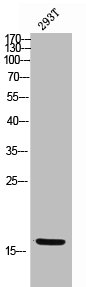
EPGN AntibodyCSB-PA006336
ApplicationsWestern Blot, ELISA
ReactivityHuman
TargetEPGN
- SizePrice
Product group Antibodies
Anti-Epigen/EPGN Antibody Picoband(r)A10011-1-CARRIER-FREE
ApplicationsWestern Blot
ReactivityHuman, Rat
TargetEPGN
- SizePrice
Product group Antibodies
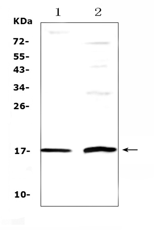
Anti-EPGN AntibodyA101453
ApplicationsWestern Blot, ELISA
ReactivityHuman
- SizePrice
Product group Antibodies

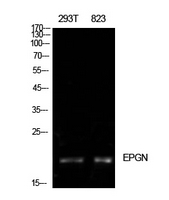
EPGN / Epigen AntibodyLS-C662272
ApplicationsWestern Blot
ReactivityHuman
TargetEPGN
- SizePrice
Product group Antibodies
References
Epigen Polyclonal AntibodyBS-5767R
ApplicationsImmunoFluorescence, Western Blot, ELISA, ImmunoCytoChemistry, ImmunoHistoChemistry, ImmunoHistoChemistry Frozen, ImmunoHistoChemistry Paraffin
ReactivityBovine, Canine, Equine, Human, Mouse, Rat, Sheep
TargetEPGN
- SizePrice
Product group Antibodies
Epigen (EPG) Polyclonal AntibodyCAU35398
ApplicationsImmunoPrecipitation, Western Blot, ImmunoCytoChemistry, ImmunoHistoChemistry
TargetEPGN
- SizePrice