


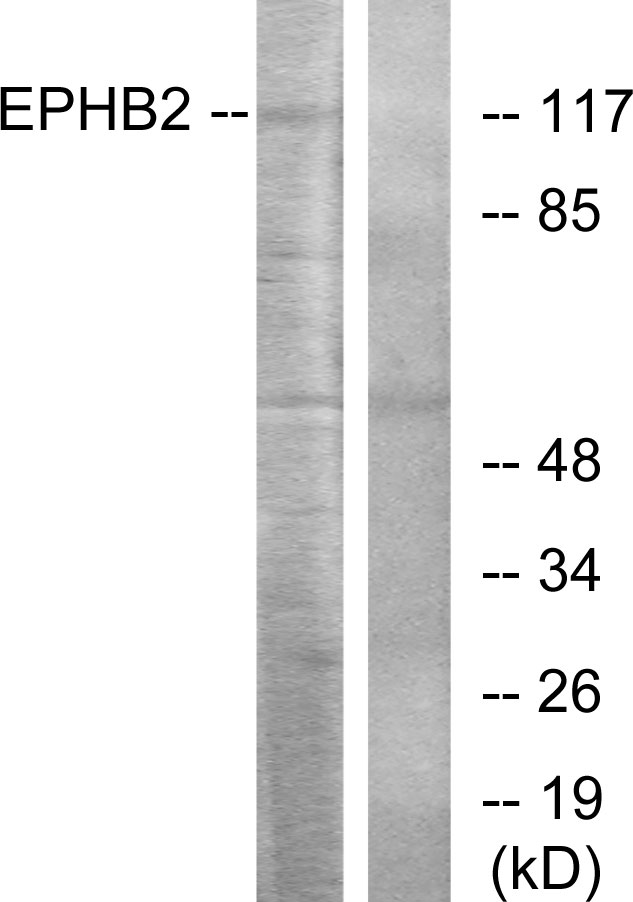
Figure 1. Western blot analysis of EPHB2 using anti-EPHB2 antibody (A01507-1). Electrophoresis was performed on a 5-20% SDS-PAGE gel at 70V (Stacking gel) / 90V (Resolving gel) for 2-3 hours. The sample well of each lane was loaded with 50ug of sample under reducing conditions. Lane 1: human U-87MG whole cell lysates, Lane 2: human A549 whole cell lysates, Lane 3: human PC-3 whole cell lysates, Lane 4: human Caco-2 whole cell lysates, Lane 5: human K562 whole cell lysates. After Electrophoresis, proteins were transferred to a Nitrocellulose membrane at 150mA for 50-90 minutes. Blocked the membrane with 5% Non-fat Milk/ TBS for 1.5 hour at RT. The membrane was incubated with rabbit anti-EPHB2 antigen affinity purified polyclonal antibody (Catalog # A01507-1) at 0.5 microg/mL overnight at 4°C, then washed with TBS-0.1%Tween 3 times with 5 minutes each and probed with a goat anti-rabbit IgG-HRP secondary antibody at a dilution of 1:5000 for 1.5 hour at RT. The signal is developed using an Enhanced Chemiluminescent detection (ECL) kit (Catalog # EK1002) with Tanon 5200 system. A specific band was detected for EPHB2 at approximately 117KD. The expected band size for EPHB2 is at 117KD.
. EPHB2 was detected in immunocytochemical section of A431 cells. Enzyme antigen retrieval was performed using IHC enzyme antigen retrieval reagent (AR0022) for 15 mins. The cells were blocked with 10% goat serum. And then incubated with 2microg/mL rabbit anti-EPHB2 Antibody (A01507-1) overnight at 4°C. DyLight®488 conjugated Goat Anti-Rabbit IgG (BA1127) was used as secondary antibody at 1:100 dilution and incubated for 30 minutes at 37°C. The section was counterstained with DAPI. Visualize using a fluorescence microscope and filter sets appropriate for the label used.")
. Overlay histogram showing A549 cells stained with A01507-1 (Blue line). To facilitate intracellular staining, cells were fixed with 4% paraformaldehyde and permeabilized with permeabilization buffer. The cells were blocked with 10% normal goat serum. And then incubated with rabbit anti-EPHB2 Antibody (A01507-1, 1microg/1x106 cells) for 30 min at 20°C. DyLight®488 conjugated goat anti-rabbit IgG (BA1127, 5-10microg/1x106 cells) was used as secondary antibody for 30 minutes at 20°C. Isotype control antibody (Green line) was rabbit IgG (1microg/1x106) used under the same conditions. Unlabelled sample without incubation with primary antibody and secondary antibody (Red line) was used as a blank control.")
. Overlay histogram showing Ana-1 cells stained with A01507-1 (Blue line). To facilitate intracellular staining, cells were fixed with 4% paraformaldehyde and permeabilized with permeabilization buffer. The cells were blocked with 10% normal goat serum. And then incubated with rabbit anti-EPHB2 Antibody (A01507-1, 1microg/1x106 cells) for 30 min at 20°C. DyLight®488 conjugated goat anti-rabbit IgG (BA1127, 5-10microg/1x106 cells) was used as secondary antibody for 30 minutes at 20°C. Isotype control antibody (Green line) was rabbit IgG (1microg/1x106) used under the same conditions. Unlabelled sample without incubation with primary antibody and secondary antibody (Red line) was used as a blank control.")
. Overlay histogram showing C6 cells stained with A01507-1 (Blue line).The cells were blocked with 10% normal goat serum. And then incubated with rabbit anti-EPHB2 Antibody (A01507-1, 1microg/1x106 cells) for 30 min at 20°C. DyLight®488 conjugated goat anti-rabbit IgG (BA1127, 5-10microg/1x106 cells) was used as secondary antibody for 30 minutes at 20°C. Isotype control antibody (Green line) was rabbit IgG (1microg/1x106) used under the same conditions. Unlabelled sample (Red line) was also used as a control.")
Figure 1. Western blot analysis of EPHB2 using anti-EPHB2 antibody (A01507-1). Electrophoresis was performed on a 5-20% SDS-PAGE gel at 70V (Stacking gel) / 90V (Resolving gel) for 2-3 hours. The sample well of each lane was loaded with 50ug of sample under reducing conditions. Lane 1: human U-87MG whole cell lysates, Lane 2: human A549 whole cell lysates, Lane 3: human PC-3 whole cell lysates, Lane 4: human Caco-2 whole cell lysates, Lane 5: human K562 whole cell lysates. After Electrophoresis, proteins were transferred to a Nitrocellulose membrane at 150mA for 50-90 minutes. Blocked the membrane with 5% Non-fat Milk/ TBS for 1.5 hour at RT. The membrane was incubated with rabbit anti-EPHB2 antigen affinity purified polyclonal antibody (Catalog # A01507-1) at 0.5 microg/mL overnight at 4°C, then washed with TBS-0.1%Tween 3 times with 5 minutes each and probed with a goat anti-rabbit IgG-HRP secondary antibody at a dilution of 1:5000 for 1.5 hour at RT. The signal is developed using an Enhanced Chemiluminescent detection (ECL) kit (Catalog # EK1002) with Tanon 5200 system. A specific band was detected for EPHB2 at approximately 117KD. The expected band size for EPHB2 is at 117KD.
Anti-Eph receptor B2/EPHB2 Antibody Picoband(r)
A01507-1-CARRIER-FREE
ApplicationsFlow Cytometry, ImmunoFluorescence, Western Blot, ELISA, ImmunoCytoChemistry
Product group Antibodies
ReactivityHuman, Mouse, Rat
TargetEPHB2
Overview
- SupplierBoster Bio
- Product NameAnti-Eph receptor B2/EPHB2 Antibody Picoband(r)
- Delivery Days Customer9
- ApplicationsFlow Cytometry, ImmunoFluorescence, Western Blot, ELISA, ImmunoCytoChemistry
- CertificationResearch Use Only
- ClonalityPolyclonal
- Concentration500 ug/ml
- Gene ID2048
- Target nameEPHB2
- Target descriptionEPH receptor B2
- Target synonymsBDPLT22, CAPB, DRT, EK5, EPHT3, ERK, Hek5, PCBC, Tyro5, ephrin type-B receptor 2, EPH-like kinase 5, developmentally-regulated Eph-related tyrosine kinase, elk-related tyrosine kinase, eph tyrosine kinase 3, protein-tyrosine kinase HEK5, renal carcinoma antigen NY-REN-47, tyrosine-protein kinase TYRO5, tyrosine-protein kinase receptor EPH-3
- HostRabbit
- IsotypeIgG
- Protein IDP29323
- Protein NameEphrin type-B receptor 2
- Scientific DescriptionBoster Bio Anti-Eph receptor B2/EPHB2 Antibody Picoband® catalog # A01507-1. Tested in ELISA, Flow Cytometry, IF, ICC, WB applications. This antibody reacts with Human, Mouse, Rat. The brand Picoband indicates this is a premium antibody that guarantees superior quality, high affinity, and strong signals with minimal background in Western blot applications. Only our best-performing antibodies are designated as Picoband, ensuring unmatched performance.
- ReactivityHuman, Mouse, Rat
- Storage Instruction-20°C,2°C to 8°C
- UNSPSC12352203
Related products
Product group Antibodies
EPHB2 AntibodyCSB-PA007730LA01HU
ApplicationsImmunoFluorescence, ELISA, ImmunoHistoChemistry
ReactivityHuman
TargetEPHB2
- SizePrice
Product group Antibodies
Anti-EPHB2 AntibodyA101110
ApplicationsWestern Blot, ELISA
ReactivityHuman
- SizePrice
Product group Antibodies

Goat anti-EPHB2 / DRT / NukEB07445
ApplicationsWestern Blot, ELISA
ReactivityCanine, Human, Mouse, Rat
TargetEPHB2
- SizePrice

Product group Antibodies
Anti-EPHB2 AntibodyHPA071200
ApplicationsWestern Blot, ImmunoCytoChemistry
ReactivityHuman
TargetEPHB2
- SizePrice
Product group Antibodies

EPHB2 / EPH Receptor B2 AntibodyLS-C411341
ApplicationsWestern Blot
ReactivityHuman, Mouse
TargetEPHB2
- SizePrice
Product group Antibodies
EphB2 antibodyGTX25418
ApplicationsImmunoHistoChemistry, ImmunoHistoChemistry Paraffin
ReactivityHuman
TargetEPHB2
- SizePrice
Product group Antibodies
EPHB2 Polyclonal AntibodyCAC13029
ApplicationsImmunoFluorescence, ELISA, ImmunoHistoChemistry
TargetEPHB2
- SizePrice
Product group Antibodies
EphB2 Polyclonal AntibodyBS-0247R
ApplicationsImmunoFluorescence, Western Blot, ELISA, ImmunoCytoChemistry, ImmunoHistoChemistry, ImmunoHistoChemistry Frozen, ImmunoHistoChemistry Paraffin
ReactivityCanine, Chicken, Human, Mouse, Rat
TargetEPHB2
- SizePrice
Product group Antibodies
Anti-EPHB2 Antibody144-09813
ApplicationsWestern Blot
ReactivityHuman, Mouse
TargetEPHB2
- SizePrice