


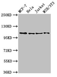
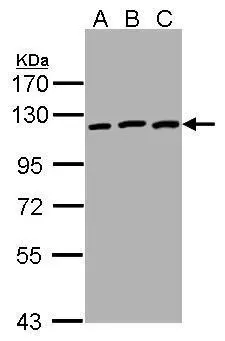
Figure 1. Western blot analysis of EPHB3 using anti-EPHB3 antibody (A04659). Electrophoresis was performed on a 5-20% SDS-PAGE gel at 70V (Stacking gel) / 90V (Resolving gel) for 2-3 hours. The sample well of each lane was loaded with 30 ug of sample under reducing conditions. Lane 1: human A431 whole cell lysates, Lane 2: human Hacat whole cell lysates, Lane 3: rat brain tissue lysates, Lane 4: rat stomach tissue lysates, Lane 5: rat lung tissue lysates, Lane 6: mouse brain tissue lysates, Lane 7: mouse stomach tissue lysates, Lane 8: mouse lung tissue lysates. After electrophoresis, proteins were transferred to a nitrocellulose membrane at 150 mA for 50-90 minutes. Blocked the membrane with 5% non-fat milk/TBS for 1.5 hour at RT. The membrane was incubated with rabbit anti-EPHB3 antigen affinity purified polyclonal antibody (Catalog # A04659) at 0.5 microg/mL overnight at 4°C, then washed with TBS-0.1%Tween 3 times with 5 minutes each and probed with a goat anti-rabbit IgG-HRP secondary antibody at a dilution of 1:5000 for 1.5 hour at RT. The signal is developed using an Enhanced Chemiluminescent detection (ECL) kit (Catalog # EK1002) with Tanon 5200 system. A specific band was detected for EPHB3 at approximately 110 kDa. The expected band size for EPHB3 is at 110-130 kDa.
Figure 1. Western blot analysis of EPHB3 using anti-EPHB3 antibody (A04659). Electrophoresis was performed on a 5-20% SDS-PAGE gel at 70V (Stacking gel) / 90V (Resolving gel) for 2-3 hours. The sample well of each lane was loaded with 30 ug of sample under reducing conditions. Lane 1: human A431 whole cell lysates, Lane 2: human Hacat whole cell lysates, Lane 3: rat brain tissue lysates, Lane 4: rat stomach tissue lysates, Lane 5: rat lung tissue lysates, Lane 6: mouse brain tissue lysates, Lane 7: mouse stomach tissue lysates, Lane 8: mouse lung tissue lysates. After electrophoresis, proteins were transferred to a nitrocellulose membrane at 150 mA for 50-90 minutes. Blocked the membrane with 5% non-fat milk/TBS for 1.5 hour at RT. The membrane was incubated with rabbit anti-EPHB3 antigen affinity purified polyclonal antibody (Catalog # A04659) at 0.5 microg/mL overnight at 4°C, then washed with TBS-0.1%Tween 3 times with 5 minutes each and probed with a goat anti-rabbit IgG-HRP secondary antibody at a dilution of 1:5000 for 1.5 hour at RT. The signal is developed using an Enhanced Chemiluminescent detection (ECL) kit (Catalog # EK1002) with Tanon 5200 system. A specific band was detected for EPHB3 at approximately 110 kDa. The expected band size for EPHB3 is at 110-130 kDa.
Anti-EPHB3 Antibody Picoband(r)
A04659-CARRIER-FREE
ApplicationsWestern Blot, ELISA, ImmunoHistoChemistry
Product group Antibodies
ReactivityHuman, Mouse, Rat
TargetEPHB3
Overview
- SupplierBoster Bio
- Product NameAnti-EPHB3 Antibody Picoband(r)
- Delivery Days Customer9
- Application Supplier NoteTested Species: In-house tested species with positive results. Other applications have not been tested. Optimal dilutions should be determined by end users.
- ApplicationsWestern Blot, ELISA, ImmunoHistoChemistry
- CertificationResearch Use Only
- ClonalityPolyclonal
- Concentration500 ug/ml
- Gene ID2049
- Target nameEPHB3
- Target descriptionEPH receptor B3
- Target synonymsEK2, ETK2, HEK2, TYRO6, ephrin type-B receptor 3, EPH-like tyrosine kinase 2, embryonic kinase 2, human embryo kinase 2, tyrosine-protein kinase TYRO6
- HostRabbit
- IsotypeIgG
- Protein IDP54753
- Protein NameEphrin type-B receptor 3
- Scientific DescriptionBoster Bio Anti-EPHB3 Antibody Picoband® catalog # A04659. Tested in ELISA, IHC, WB applications. This antibody reacts with Human, Mouse, Rat. The brand Picoband indicates this is a premium antibody that guarantees superior quality, high affinity, and strong signals with minimal background in Western blot applications. Only our best-performing antibodies are designated as Picoband, ensuring unmatched performance.
- ReactivityHuman, Mouse, Rat
- Storage Instruction-20°C,2°C to 8°C
- UNSPSC12352203
Related products
Product group Antibodies
ApplicationsWestern Blot
ReactivityHuman, Mouse, Rat
- SizePrice
Product group Antibodies
Anti-Ephb3 (Center) Antibody102-24175
ApplicationsWestern Blot
TargetEPHB3
- SizePrice
Product group Antibodies
ApplicationsFlow Cytometry, Western Blot
ReactivityHuman, Mouse, Rat
TargetEPHB3
- SizePrice
Product group Antibodies
EPHB3 AntibodyCSB-PA007731LA01HU
ApplicationsImmunoFluorescence, Western Blot, ELISA
ReactivityHuman, Mouse, Rat
TargetEPHB3
- SizePrice
Product group Antibodies
Ephb3 Polyclonal AntibodyCAC11723
ApplicationsImmunoFluorescence, Western Blot, ELISA
ReactivityMouse, Rat
TargetEPHB3
- SizePrice
Product group Antibodies

EPHB3 / EPH Receptor B3 AntibodyLS-C402657
ApplicationsELISA, ImmunoHistoChemistry
ReactivityHuman, Mouse
TargetEPHB3
- SizePrice

Product group Antibodies
Anti-EPHB3 AntibodyHPA008184
ApplicationsWestern Blot, ImmunoHistoChemistry
ReactivityHuman
TargetEPHB3
- SizePrice
Product group Antibodies
EphB3 antibody [C1C3]GTX107882
ApplicationsWestern Blot
ReactivityHuman
TargetEPHB3
- SizePrice