

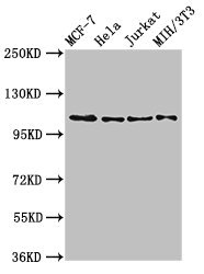
Western Blot Positive WB detected in: MCF-7 whole cell lysate, Hela whole cell lysate, Jurkat whole cell lysate, NIH/3T3 whole cell lysate All lanes: EPHB3 antibody at 2.7ug/ml Secondary Goat polyclonal to rabbit IgG at 1/50000 dilution Predicted band size: 111 kDa Observed band size: 111 kDa
")
Western Blot Positive WB detected in: MCF-7 whole cell lysate, Hela whole cell lysate, Jurkat whole cell lysate, NIH/3T3 whole cell lysate All lanes: EPHB3 antibody at 2.7ug/ml Secondary Goat polyclonal to rabbit IgG at 1/50000 dilution Predicted band size: 111 kDa Observed band size: 111 kDa
EPHB3 Antibody
CSB-PA007731LA01HU
ApplicationsImmunoFluorescence, Western Blot, ELISA
Product group Antibodies
ReactivityHuman, Mouse, Rat
TargetEPHB3
Overview
- SupplierCusabio
- Product NameEPHB3 Antibody
- Delivery Days Customer20
- ApplicationsImmunoFluorescence, Western Blot, ELISA
- CertificationResearch Use Only
- ClonalityPolyclonal
- ConjugateUnconjugated
- Gene ID2049
- Target nameEPHB3
- Target descriptionEPH receptor B3
- Target synonymsEK2, ETK2, HEK2, TYRO6, ephrin type-B receptor 3, EPH-like tyrosine kinase 2, embryonic kinase 2, human embryo kinase 2, tyrosine-protein kinase TYRO6
- HostRabbit
- IsotypeIgG
- Protein IDP54753
- Protein NameEphrin type-B receptor 3
- Scientific DescriptionReceptor tyrosine kinase which binds promiscuously transmembrane ephrin-B family ligands residing on adjacent cells, leading to contact-dependent bidirectional signaling into neighboring cells. The signaling pathway downstream of the receptor is referred to as forward signaling while the signaling pathway downstream of the ephrin ligand is referred to as reverse signaling. Generally has an overlapping and redundant function with EPHB2. Like EPHB2, functions in axon guidance during development regulating for instance the neurons forming the corpus callosum and the anterior commissure, 2 major interhemispheric connections between the temporal lobes of the cerebral cortex. In addition to its role in axon guidance plays also an important redundant role with other ephrin-B receptors in development and maturation of dendritic spines and the formation of excitatory synapses. Controls other aspects of development through regulation of cell migration and positioning. This includes angiogenesis, palate development and thymic epithelium development for instance. Forward and reverse signaling through the EFNB2/EPHB3 complex also regulate migration and adhesion of cells that tubularize the urethra and septate the cloaca. Finally, plays an important role in intestinal epithelium differentiation segregating progenitor from differentiated cells in the crypt.
- ReactivityHuman, Mouse, Rat
- Storage Instruction-20°C or -80°C
- UNSPSC41116161
Related products
Product group Antibodies
ApplicationsWestern Blot
ReactivityHuman, Mouse, Rat
- SizePrice
Product group Antibodies
Anti-Ephb3 (Center) Antibody102-24175
ApplicationsWestern Blot
TargetEPHB3
- SizePrice

Product group Antibodies
Anti-EPHB3 Antibody Picoband(r)A04659-CARRIER-FREE
ApplicationsWestern Blot, ELISA, ImmunoHistoChemistry
ReactivityHuman, Mouse, Rat
TargetEPHB3
- SizePrice
Product group Antibodies
ApplicationsFlow Cytometry, Western Blot
ReactivityHuman, Mouse, Rat
TargetEPHB3
- SizePrice
Product group Antibodies
Ephb3 Polyclonal AntibodyCAC11723
ApplicationsImmunoFluorescence, Western Blot, ELISA
ReactivityMouse, Rat
TargetEPHB3
- SizePrice
Product group Antibodies

EPHB3 / EPH Receptor B3 AntibodyLS-C402657
ApplicationsELISA, ImmunoHistoChemistry
ReactivityHuman, Mouse
TargetEPHB3
- SizePrice

Product group Antibodies
Anti-EPHB3 AntibodyHPA008184
ApplicationsWestern Blot, ImmunoHistoChemistry
ReactivityHuman
TargetEPHB3
- SizePrice
Product group Antibodies
EphB3 antibody [C1C3]GTX107882
ApplicationsWestern Blot
ReactivityHuman
TargetEPHB3
- SizePrice