


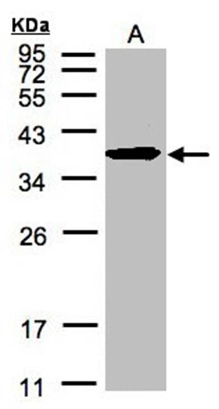
Figure 1. Western blot analysis of Erlin-2/ERLIN2 using anti-Erlin-2/ERLIN2 antibody (A07042-2). Electrophoresis was performed on a 5-20% SDS-PAGE gel at 70V (Stacking gel) / 90V (Resolving gel) for 2-3 hours. The sample well of each lane was loaded with 30 ug of sample under reducing conditions. Lane 1: human HepG2 whole cell lysates, Lane 2: human Hela whole cell lysates, Lane 3: human T-47D whole cell lysates, Lane 4: human U-87MG whole cell lysates, Lane 5: rat kidney tissue lysates, Lane 6: rat testis tissue lysates, Lane 8: rat liver tissue lysates, Lane 9: mouse kidney tissue lysates, Lane 10: mouse testis tissue lysates. After electrophoresis, proteins were transferred to a nitrocellulose membrane at 150 mA for 50-90 minutes. Blocked the membrane with 5% non-fat milk/TBS for 1.5 hour at RT. The membrane was incubated with rabbit anti-Erlin-2/ERLIN2 antigen affinity purified polyclonal antibody (Catalog # A07042-2) at 0.25 microg/mL overnight at 4°C, then washed with TBS-0.1%Tween 3 times with 5 minutes each and probed with a goat anti-rabbit IgG-HRP secondary antibody at a dilution of 1:5000 for 1.5 hour at RT. The signal is developed using an Enhanced Chemiluminescent detection (ECL) kit (Catalog # EK1002) with Tanon 5200 system. A specific band was detected for Erlin-2/ERLIN2 at approximately 43 kDa. The expected band size for Erlin-2/ERLIN2 is at 43 kDa.
. Erlin-2/ERLIN2 was detected in an immunocytochemical section of T-47D cells. Enzyme antigen retrieval was performed using IHC enzyme antigen retrieval reagent (AR0022) for 15 mins. The cells were blocked with 10% goat serum. And then incubated with 5 microg/mL rabbit anti-Erlin-2/ERLIN2 Antibody (A07042-2) overnight at 4°C. DyLight®488 Conjugated Goat Anti-Rabbit IgG (BA1127) was used as secondary antibody at 1:100 dilution and incubated for 30 minutes at 37°C. The section was counterstained with DAPI. Visualize using a fluorescence microscope and filter sets appropriate for the label used.")
. Overlay histogram showing Caco-2 cells stained with A07042-2 (Blue line). To facilitate intracellular staining, cells were fixed with 4% paraformaldehyde and permeabilized with permeabilization buffer. The cells were blocked with 10% normal goat serum. And then incubated with rabbit anti-Erlin-2/ERLIN2 Antibody (A07042-2, 1 microg/1x106 cells) for 30 min at 20°C. DyLight®488 conjugated goat anti-rabbit IgG (BA1127, 5-10 microg/1x106 cells) was used as secondary antibody for 30 minutes at 20°C. Isotype control antibody (Green line) was rabbit IgG (1 microg/1x106) used under the same conditions. Unlabelled sample without incubation with primary antibody and secondary antibody (Red line) was used as a blank control.")
Figure 1. Western blot analysis of Erlin-2/ERLIN2 using anti-Erlin-2/ERLIN2 antibody (A07042-2). Electrophoresis was performed on a 5-20% SDS-PAGE gel at 70V (Stacking gel) / 90V (Resolving gel) for 2-3 hours. The sample well of each lane was loaded with 30 ug of sample under reducing conditions. Lane 1: human HepG2 whole cell lysates, Lane 2: human Hela whole cell lysates, Lane 3: human T-47D whole cell lysates, Lane 4: human U-87MG whole cell lysates, Lane 5: rat kidney tissue lysates, Lane 6: rat testis tissue lysates, Lane 8: rat liver tissue lysates, Lane 9: mouse kidney tissue lysates, Lane 10: mouse testis tissue lysates. After electrophoresis, proteins were transferred to a nitrocellulose membrane at 150 mA for 50-90 minutes. Blocked the membrane with 5% non-fat milk/TBS for 1.5 hour at RT. The membrane was incubated with rabbit anti-Erlin-2/ERLIN2 antigen affinity purified polyclonal antibody (Catalog # A07042-2) at 0.25 microg/mL overnight at 4°C, then washed with TBS-0.1%Tween 3 times with 5 minutes each and probed with a goat anti-rabbit IgG-HRP secondary antibody at a dilution of 1:5000 for 1.5 hour at RT. The signal is developed using an Enhanced Chemiluminescent detection (ECL) kit (Catalog # EK1002) with Tanon 5200 system. A specific band was detected for Erlin-2/ERLIN2 at approximately 43 kDa. The expected band size for Erlin-2/ERLIN2 is at 43 kDa.
Anti-Erlin-2/ERLIN2 Antibody Picoband(r)
A07042-2-CARRIER-FREE
ApplicationsFlow Cytometry, ImmunoFluorescence, Western Blot, ELISA, ImmunoCytoChemistry
Product group Antibodies
ReactivityHuman, Mouse, Rat
TargetERLIN2
Overview
- SupplierBoster Bio
- Product NameAnti-Erlin-2/ERLIN2 Antibody Picoband(r)
- Delivery Days Customer9
- ApplicationsFlow Cytometry, ImmunoFluorescence, Western Blot, ELISA, ImmunoCytoChemistry
- CertificationResearch Use Only
- ClonalityPolyclonal
- Concentration500 ug/ml
- Gene ID11160
- Target nameERLIN2
- Target descriptionER lipid raft associated 2
- Target synonymsC8orf2, Erlin-2, NET32, SPFH2, SPG18, SPG18A, SPG18B, erlin-2, SPFH domain family, member 2, endoplasmic reticulum lipid raft-associated protein 2, epididymis secretory sperm binding protein, spastic paraplegia 18 (autosomal dominant), stomatin-prohibitin-flotillin-HflC/K domain-containing protein 2
- HostRabbit
- IsotypeIgG
- Protein IDO94905
- Protein NameErlin-2
- Scientific DescriptionBoster Bio Anti-Erlin-2/ERLIN2 Antibody Picoband® catalog # A07042-2. Tested in ELISA, Flow Cytometry, IF, ICC, WB applications. This antibody reacts with Human, Mouse, Rat. The brand Picoband indicates this is a premium antibody that guarantees superior quality, high affinity, and strong signals with minimal background in Western blot applications. Only our best-performing antibodies are designated as Picoband, ensuring unmatched performance.
- ReactivityHuman, Mouse, Rat
- Storage Instruction-20°C,2°C to 8°C
- UNSPSC12352203
Related products
Product group Antibodies
ERLIN2 AntibodyCSB-PA007791ESR1HU
ApplicationsELISA, ImmunoHistoChemistry
ReactivityHuman
TargetERLIN2
- SizePrice
Product group Antibodies
Anti-SPFH2 AntibodyA37479
ApplicationsWestern Blot, ImmunoHistoChemistry
ReactivityHuman
- SizePrice
Product group Antibodies

Goat anti-SPFH2 / ERLIN2EB06896
ApplicationsWestern Blot, ELISA, ImmunoHistoChemistry
ReactivityHuman, Rat
TargetERLIN2
- SizePrice

Product group Antibodies
Anti-ERLIN2 AntibodyHPA002025
ApplicationsWestern Blot, ImmunoCytoChemistry, ImmunoHistoChemistry
ReactivityHuman, Mouse, Rat
TargetERLIN2
- SizePrice
Product group Antibodies

SPFH2 / ERLIN2 AntibodyLS-C409849
ApplicationsWestern Blot
ReactivityHuman
TargetERLIN2
- SizePrice
Product group Antibodies
ERLIN2 Polyclonal AntibodyCAC13031
ApplicationsWestern Blot, ELISA, ImmunoHistoChemistry
TargetERLIN2
- SizePrice
Product group Antibodies
SPFH2 antibody [C2C3], C-termGTX106277
ApplicationsWestern Blot, ImmunoHistoChemistry, ImmunoHistoChemistry Paraffin
ReactivityHuman
TargetERLIN2
- SizePrice
Product group Antibodies
Anti-ERLIN2 (C-term) Antibody102-23242
ApplicationsWestern Blot
TargetERLIN2
- SizePrice
Product group Antibodies
SPFH2 Recombinant AntibodyBSM-62469R
ApplicationsImmunoFluorescence, Western Blot, ImmunoCytoChemistry, ImmunoHistoChemistry, ImmunoHistoChemistry Frozen, ImmunoHistoChemistry Paraffin
ReactivityHuman, Mouse
TargetERLIN2
- SizePrice