


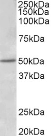
Figure 1. Western blot analysis of ESR2 using anti-ESR2 antibody (A00786-1). Electrophoresis was performed on a 5-20% SDS-PAGE gel at 70V (Stacking gel) / 90V (Resolving gel) for 2-3 hours. The sample well of each lane was loaded with 30 ug of sample under reducing conditions. Lane 1: human PC-3 whole cell lysates, Lane 2: human MCF-7 whole cell lysates, Lane 3: rat brain tissue lysates, Lane 4: rat testis tissue lysates, Lane 5: mouse brain tissue lysates, Lane 6: mouse testis tissue lysates, Lane 7: mouse NIH/3T3 whole cell lysates. After electrophoresis, proteins were transferred to a nitrocellulose membrane at 150 mA for 50-90 minutes. Blocked the membrane with 5% non-fat milk/TBS for 1.5 hour at RT. The membrane was incubated with rabbit anti-ESR2 antigen affinity purified polyclonal antibody (Catalog # A00786-1) at 0.5 microg/mL overnight at 4°C, then washed with TBS-0.1%Tween 3 times with 5 minutes each and probed with a goat anti-rabbit IgG-HRP secondary antibody at a dilution of 1:5000 for 1.5 hour at RT. The signal is developed using an Enhanced Chemiluminescent detection (ECL) kit (Catalog # EK1002) with Tanon 5200 system. A specific band was detected for ESR2 at approximately 59 kDa. The expected band size for ESR2 is at 59 kDa.
Figure 1. Western blot analysis of ESR2 using anti-ESR2 antibody (A00786-1). Electrophoresis was performed on a 5-20% SDS-PAGE gel at 70V (Stacking gel) / 90V (Resolving gel) for 2-3 hours. The sample well of each lane was loaded with 30 ug of sample under reducing conditions. Lane 1: human PC-3 whole cell lysates, Lane 2: human MCF-7 whole cell lysates, Lane 3: rat brain tissue lysates, Lane 4: rat testis tissue lysates, Lane 5: mouse brain tissue lysates, Lane 6: mouse testis tissue lysates, Lane 7: mouse NIH/3T3 whole cell lysates. After electrophoresis, proteins were transferred to a nitrocellulose membrane at 150 mA for 50-90 minutes. Blocked the membrane with 5% non-fat milk/TBS for 1.5 hour at RT. The membrane was incubated with rabbit anti-ESR2 antigen affinity purified polyclonal antibody (Catalog # A00786-1) at 0.5 microg/mL overnight at 4°C, then washed with TBS-0.1%Tween 3 times with 5 minutes each and probed with a goat anti-rabbit IgG-HRP secondary antibody at a dilution of 1:5000 for 1.5 hour at RT. The signal is developed using an Enhanced Chemiluminescent detection (ECL) kit (Catalog # EK1002) with Tanon 5200 system. A specific band was detected for ESR2 at approximately 59 kDa. The expected band size for ESR2 is at 59 kDa.
Anti-Estrogen Receptor beta/ESR2 Antibody Picoband(r)
A00786-1-CARRIER-FREE
ApplicationsWestern Blot
Product group Antibodies
ReactivityHuman, Mouse, Rat
TargetESR2
Overview
- SupplierBoster Bio
- Product NameAnti-Estrogen Receptor beta/ESR2 Antibody Picoband(r)
- Delivery Days Customer9
- ApplicationsWestern Blot
- CertificationResearch Use Only
- ClonalityPolyclonal
- Concentration500 ug/ml
- Gene ID2100
- Target nameESR2
- Target descriptionestrogen receptor 2
- Target synonymsER-BETA, ESR-BETA, ESRB, ESTRB, Erb, NR3A2, ODG8, estrogen receptor beta, estrogen receptor beta 2, estrogen receptor beta 4, nuclear receptor subfamily 3 group A member 2, oestrogen receptor beta
- HostRabbit
- IsotypeIgG
- Protein IDQ92731
- Protein NameEstrogen receptor beta
- Scientific DescriptionBoster Bio Anti-Estrogen Receptor beta/ESR2 Antibody Picoband® catalog # A00786-1. Tested in WB applications. This antibody reacts with Human, Mouse, Rat. The brand Picoband indicates this is a premium antibody that guarantees superior quality, high affinity, and strong signals with minimal background in Western blot applications. Only our best-performing antibodies are designated as Picoband, ensuring unmatched performance.
- ReactivityHuman, Mouse, Rat
- Storage Instruction-20°C,2°C to 8°C
- UNSPSC12352203
Related products
Product group Antibodies
ApplicationsImmunoFluorescence, Western Blot, ELISA
ReactivityHuman
- SizePrice
Product group Antibodies

Anti-estrogen receptor beta [CO1531]AB02568-10.0-BT
ApplicationsImmunoPrecipitation, Western Blot
ReactivityHuman
TargetESR2
- SizePrice
Product group Antibodies
Anti-ESR2 Antibody144-02546
ApplicationsWestern Blot, ImmunoHistoChemistry
ReactivityHuman, Mouse, Rat
TargetESR2
- SizePrice
Product group Antibodies
References
Estrogen receptor beta AntibodyBS-0116R
ApplicationsImmunoFluorescence, Western Blot, ELISA, ImmunoCytoChemistry, ImmunoHistoChemistry, ImmunoHistoChemistry Frozen, ImmunoHistoChemistry Paraffin
ReactivityHuman, Mouse, Rat
TargetESR2
- SizePrice
Product group Antibodies
ESR2 AntibodyCSB-PA002446
ApplicationsImmunoFluorescence, Western Blot, ELISA
ReactivityHuman, Mouse, Rat
TargetESR2
- SizePrice
Product group Antibodies

ApplicationsWestern Blot, ELISA, ImmunoHistoChemistry
ReactivityHuman
TargetESR2
- SizePrice
Product group Antibodies
Esr2 Polyclonal AntibodyCAC07131
ApplicationsWestern Blot, ELISA, ImmunoHistoChemistry
ReactivityMouse
TargetESR2
- SizePrice
Product group Antibodies
Estrogen Receptor beta antibodyGTX134663
ApplicationsWestern Blot
ReactivityHuman, Mouse
TargetESR2
- SizePrice
Product group Antibodies

ESR2 / ER Beta Antibody (phospho-Ser105)LS-C358984
ApplicationsImmunoFluorescence, Western Blot, ImmunoCytoChemistry
ReactivityCanine, Human, Mouse, Rat
TargetESR2
- SizePrice