


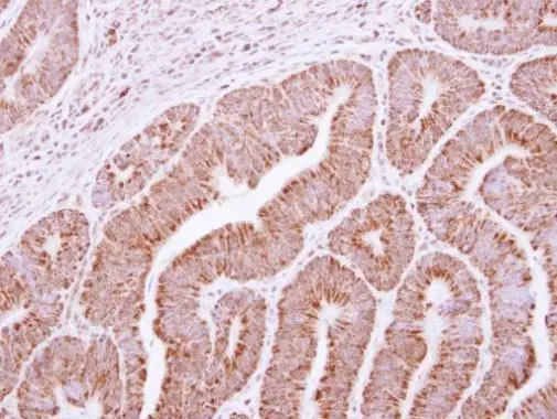
Immunohistochemical staining of human kidney shows strong positivity in tubules.
![Lane 1: Marker [kDa] 230, 130, 95, 72, 56, 36, 28, 17, 11. Lane 2: Human cell line RT-4. Lane 3: Human cell line U-251MG sp](https://atlasantibodies.s3.amazonaws.com/images/wb/hpa021790-wb-1.jpg "Lane 1: Marker [kDa] 230, 130, 95, 72, 56, 36, 28, 17, 11. Lane 2: Human cell line RT-4. Lane 3: Human cell line U-251MG sp")
Immunohistochemical staining of human kidney shows strong positivity in tubules.
Anti-EXOSC2 Antibody
HPA021790
ApplicationsWestern Blot, ImmunoHistoChemistry
Product group Antibodies
ReactivityHuman, Mouse, Rat
TargetEXOSC2
Overview
- SupplierAtlas Antibodies
- Product NameAnti-EXOSC2 Antibody
- Delivery Days Customer4
- ApplicationsWestern Blot, ImmunoHistoChemistry
- CertificationResearch Use Only
- ClonalityPolyclonal
- ConjugateUnconjugated
- Gene ID23404
- Target nameEXOSC2
- Target descriptionexosome component 2
- Target synonymsRRP4, Rrp4p, SHRF, hRrp4p, p7, exosome complex component RRP4, exosome complex exonuclease RRP4, homolog of yeast RRP4 (ribosomal RNA processing 4), 3' 5' exoribonuclease (RRP4), homolog of yeast RRP4 (ribosomal RNA processing 4), 3'-5'-exoribonuclease, ribosomal RNA-processing protein 4
- HostRabbit
- IsotypeIgG
- Protein IDQ13868
- Protein NameExosome complex component RRP4
- Scientific DescriptionRecombinant Protein Epitope Signature Tag (PrEST) antigen sequence
- ReactivityHuman, Mouse, Rat
- Storage Instruction-20°C,2°C to 8°C
- UNSPSC41116161
Datasheet
MSDS
Related products

Product group Antibodies
Anti-RRP4/EXOSC2 Antibody Picoband(r)A10119-1-CARRIER-FREE
ApplicationsFlow Cytometry, Western Blot, ELISA, ImmunoHistoChemistry
ReactivityHuman
TargetEXOSC2
- SizePrice
Product group Antibodies
Anti-EXOSC2 Antibody144-10450
ApplicationsWestern Blot
ReactivityHuman, Mouse, Rat
TargetEXOSC2
- SizePrice
Product group Antibodies
EXOSC2 AntibodyCSB-PA007890GA01HU
ApplicationsImmunoFluorescence, Western Blot, ELISA, ImmunoHistoChemistry
ReactivityHuman, Mouse, Rat
TargetEXOSC2
- SizePrice
Product group Antibodies
ApplicationsImmunoPrecipitation, Western Blot, ImmunoCytoChemistry, ImmunoHistoChemistry
TargetEXOSC2
- SizePrice
Product group Antibodies

EXOSC2 / RRP4 AntibodyLS-C497159
ApplicationsWestern Blot
ReactivityHuman, Mouse, Rat
TargetEXOSC2
- SizePrice

Product group Antibodies
Anti-EXOSC2 AntibodyHPA021756
ApplicationsImmunoCytoChemistry
ReactivityHuman
TargetEXOSC2
- SizePrice
Product group Antibodies
Anti-EXOSC2 AntibodyHPA021756
ApplicationsImmunoCytoChemistry
ReactivityHuman
TargetEXOSC2
- SizePrice
Product group Antibodies
RRP4 antibody [N1C3]GTX117616
ApplicationsImmunoFluorescence, Western Blot, ImmunoCytoChemistry, ImmunoHistoChemistry, ImmunoHistoChemistry Paraffin
ReactivityHuman
TargetEXOSC2
- SizePrice