


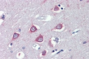
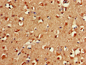
Immunohistochemical staining of human Prostate shows strong cytoplasmic positivity in glandular cells.
Immunohistochemical staining of human Prostate shows strong cytoplasmic positivity in glandular cells.
Anti-FAAH Antibody
HPA007425
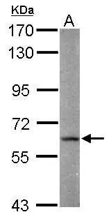
ApplicationsImmunoHistoChemistry
Product group Antibodies
ReactivityHuman
TargetFAAH
Overview
- SupplierAtlas Antibodies
- Product NameAnti-FAAH Antibody
- Delivery Days Customer4
- ApplicationsImmunoHistoChemistry
- CertificationResearch Use Only
- ClonalityPolyclonal
- ConjugateUnconjugated
- Gene ID2166
- Target nameFAAH
- Target descriptionfatty acid amide hydrolase
- Target synonymsFAAH-1, FAAH1, PSAB, fatty-acid amide hydrolase 1, anandamide amidohydrolase 1, fatty acid ester hydrolase, oleamide hydrolase 1
- HostRabbit
- IsotypeIgG
- Protein IDO00519
- Protein NameFatty-acid amide hydrolase 1
- Scientific DescriptionRecombinant Protein Epitope Signature Tag (PrEST) antigen sequence
- ReactivityHuman
- Storage Instruction-20°C,2°C to 8°C
- UNSPSC41116161
Datasheet
MSDS
Related products

Product group Antibodies
Anti-FAAH1/FAAH Antibody Picoband(r)A00801-2-CARRIER-FREE
ApplicationsFlow Cytometry, ImmunoFluorescence, Western Blot, ELISA, ImmunoCytoChemistry
ReactivityHuman, Mouse, Rat
TargetFAAH
- SizePrice
Product group Antibodies
Anti-FAAH Antibody144-01174
ApplicationsWestern Blot
ReactivityHuman, Mouse, Rat
TargetFAAH
- SizePrice
Product group Antibodies
Anti-FAAH1 AntibodyA326243
ApplicationsELISA, ImmunoHistoChemistry
ReactivityHuman
- SizePrice

Product group Antibodies
FAAH (9C3) Monoclonal AntibodyBSM-54433R
ApplicationsWestern Blot, ImmunoHistoChemistry, ImmunoHistoChemistry Paraffin
ReactivityHuman, Mouse
TargetFAAH
- SizePrice
Product group Antibodies
FAAH AntibodyCSB-PA007938LA01HU
ApplicationsImmunoFluorescence, ELISA, ImmunoHistoChemistry
ReactivityHuman
TargetFAAH
- SizePrice
Product group Antibodies

Goat anti-FAAHEB08851
ApplicationsELISA, ImmunoHistoChemistry
ReactivityCanine, Human, Mouse, Rat
TargetFAAH
- SizePrice
Product group Antibodies
Faah Recombinant AntibodyCAC12165
ApplicationsWestern Blot, ELISA, ImmunoHistoChemistry
ReactivityMouse
TargetFAAH
- SizePrice
Product group Antibodies

FAAH AntibodyLS-C401654
ApplicationsELISA, ImmunoHistoChemistry
ReactivityHuman, Mouse, Rat
TargetFAAH
- SizePrice
Product group Antibodies
FAAH antibody [C2C3], C-termGTX124189
ApplicationsWestern Blot, ImmunoHistoChemistry, ImmunoHistoChemistry Paraffin
ReactivityHuman
TargetFAAH
- SizePrice