


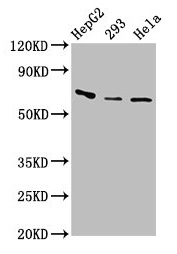
Figure 1. Western blot analysis of Factor I using anti-Factor I antibody (PB9935). Electrophoresis was performed on a 5-20% SDS-PAGE gel at 70V (Stacking gel) / 90V (Resolving gel) for 2-3 hours. The sample well of each lane was loaded with 50ug of sample under reducing conditions. Lane 1: rat liver tissue lysates, Lane 2: HELA whole cell lysates. After Electrophoresis, proteins were transferred to a Nitrocellulose membrane at 150mA for 50-90 minutes. Blocked the membrane with 5% Non-fat Milk/ TBS for 1.5 hour at RT. The membrane was incubated with rabbit anti-Factor I antigen affinity purified polyclonal antibody (Catalog # PB9935) at 0.5 microg/mL overnight at 4°C, then washed with TBS-0.1%Tween 3 times with 5 minutes each and probed with a goat anti-rabbit IgG-HRP secondary antibody at a dilution of 1:10000 for 1.5 hour at RT. The signal is developed using an Enhanced Chemiluminescent detection (ECL) kit (Catalog # EK1002) with Tanon 5200 system. A specific band was detected for Factor I at approximately 75KD; 45KD. The expected band size for Factor I is at 66KD.
. Overlay histogram showing U-87 cells stained with PB9935 (Blue line). To facilitate intracellular staining, cells were fixed with 4% paraformaldehyde and permeabilized with permeabilization buffer. The cells were blocked with 10% normal goat serum. And then incubated with rabbit anti-Factor I Antibody (PB9935,1microg/1x106 cells) for 30 min at 20°C. DyLight®488 conjugated goat anti-rabbit IgG (BA1127, 5-10microg/1x106 cells) was used as secondary antibody for 30 minutes at 20°C. Isotype control antibody (Green line) was rabbit IgG (1microg/1x106) used under the same conditions. Unlabelled sample without incubation with primary antibody and secondary antibody (Red line) was used as a blank control.")
. Overlay histogram showing HEPG2 cells stained with PB9935 (Blue line). To facilitate intracellular staining, cells were fixed with 4% paraformaldehyde and permeabilized with permeabilization buffer. The cells were blocked with 10% normal goat serum. And then incubated with rabbit anti-Factor I Antibody (PB9935,1microg/1x106 cells) for 30 min at 20°C. DyLight®488 conjugated goat anti-rabbit IgG (BA1127, 5-10microg/1x106 cells) was used as secondary antibody for 30 minutes at 20°C. Isotype control antibody (Green line) was rabbit IgG (1microg/1x106) used under the same conditions. Unlabelled sample without incubation with primary antibody and secondary antibody (Red line) was used as a blank control.")
Figure 1. Western blot analysis of Factor I using anti-Factor I antibody (PB9935). Electrophoresis was performed on a 5-20% SDS-PAGE gel at 70V (Stacking gel) / 90V (Resolving gel) for 2-3 hours. The sample well of each lane was loaded with 50ug of sample under reducing conditions. Lane 1: rat liver tissue lysates, Lane 2: HELA whole cell lysates. After Electrophoresis, proteins were transferred to a Nitrocellulose membrane at 150mA for 50-90 minutes. Blocked the membrane with 5% Non-fat Milk/ TBS for 1.5 hour at RT. The membrane was incubated with rabbit anti-Factor I antigen affinity purified polyclonal antibody (Catalog # PB9935) at 0.5 microg/mL overnight at 4°C, then washed with TBS-0.1%Tween 3 times with 5 minutes each and probed with a goat anti-rabbit IgG-HRP secondary antibody at a dilution of 1:10000 for 1.5 hour at RT. The signal is developed using an Enhanced Chemiluminescent detection (ECL) kit (Catalog # EK1002) with Tanon 5200 system. A specific band was detected for Factor I at approximately 75KD; 45KD. The expected band size for Factor I is at 66KD.
Anti-Factor I/CFI Antibody Picoband(r)
PB9935-CARRIER-FREE
ApplicationsFlow Cytometry, Western Blot, ImmunoCytoChemistry, ImmunoHistoChemistry
Product group Antibodies
ReactivityHuman, Rat
TargetCFI
Overview
- SupplierBoster Bio
- Product NameAnti-Factor I/CFI Antibody Picoband(r)
- Delivery Days Customer9
- Application Supplier NoteTested Species: In-house tested species with positive results. Other applications have not been tested. Optimal dilutions should be determined by end users.
- ApplicationsFlow Cytometry, Western Blot, ImmunoCytoChemistry, ImmunoHistoChemistry
- CertificationResearch Use Only
- ClonalityPolyclonal
- Concentration500 ug/ml
- Gene ID3426
- Target nameCFI
- Target descriptioncomplement factor I
- Target synonymsAHUS3, ARMD13, C3BINA, C3b-INA, FI, IF, KAF, complement factor I, C3B/C4B inactivator, C3b-inactivator, Konglutinogen-activating factor, complement component I, complement control protein factor I, complement factor I heavy chain, light chain of factor I
- HostRabbit
- IsotypeIgG
- Protein IDP05156
- Protein NameComplement factor I
- Scientific DescriptionBoster Bio Anti-Factor I/CFI Antibody Picoband® catalog # PB9935. Tested in Flow Cytometry, IHC, ICC, WB applications. This antibody reacts with Human, Rat. The brand Picoband indicates this is a premium antibody that guarantees superior quality, high affinity, and strong signals with minimal background in Western blot applications. Only our best-performing antibodies are designated as Picoband, ensuring unmatched performance.
- ReactivityHuman, Rat
- Storage Instruction-20°C,2°C to 8°C
- UNSPSC12352203
Related products
Product group Antibodies
CFI AntibodyCSB-PA005279LA01HU
ApplicationsWestern Blot, ELISA, ImmunoHistoChemistry
ReactivityHuman
TargetCFI
- SizePrice
Product group Antibodies
Anti-CFI AntibodyA101638
ApplicationsWestern Blot, ELISA
ReactivityHuman
- SizePrice
Product group Antibodies
Anti-CFI Antibody144-05623
ApplicationsImmunoFluorescence, Western Blot
ReactivityHuman, Mouse
TargetCFI
- SizePrice
Product group Antibodies

Anti-Complement Factor I [OX-21]AB00565-1.1-BT
ApplicationsFlow Cytometry, ImmunoPrecipitation, Western Blot, ELISA, ImmunoHistoChemistry, ImmunoHistoChemistry Paraffin, RadioImmunoAssay
ReactivityHuman
TargetCFI
- SizePrice

Product group Antibodies
Anti-CFI AntibodyHPA001143
ApplicationsImmunoHistoChemistry
ReactivityHuman
TargetCFI
- SizePrice
Product group Antibodies

CFI / Complement Factor I AntibodyLS-C346093
ApplicationsImmunoFluorescence, Western Blot
ReactivityHuman, Mouse
TargetCFI
- SizePrice


Product group Antibodies
Factor I antibodyGTX32531
ApplicationsWestern Blot, ImmunoHistoChemistry, ImmunoHistoChemistry Paraffin
ReactivityHuman
TargetCFI
- SizePrice
Product group Antibodies
ApplicationsImmunoFluorescence, Western Blot, ELISA, ImmunoCytoChemistry, ImmunoHistoChemistry, ImmunoHistoChemistry Frozen, ImmunoHistoChemistry Paraffin
ReactivityBovine, Equine, Human, Mouse, Rat
TargetCFI
- SizePrice