


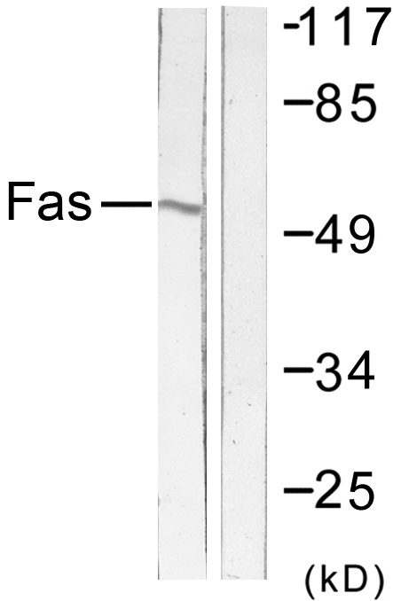
Figure 1. Western blot analysis of Fas using anti-Fas antibody (A00055). Electrophoresis was performed on a 5-20% SDS-PAGE gel at 70V (Stacking gel) / 90V (Resolving gel) for 2-3 hours. The sample well of each lane was loaded with 50ug of sample under reducing conditions. Lane 1: rat thymus tissue lysates, Lane 2: mouse RAW264.7 whole cell lysates. After Electrophoresis, proteins were transferred to a Nitrocellulose membrane at 150mA for 50-90 minutes. Blocked the membrane with 5% Non-fat Milk/ TBS for 1.5 hour at RT. The membrane was incubated with rabbit anti-Fas antigen affinity purified polyclonal antibody (Catalog # A00055) at 0.5 microg/mL overnight at 4°C, then washed with TBS-0.1%Tween 3 times with 5 minutes each and probed with a goat anti-rabbit IgG-HRP secondary antibody at a dilution of 1:5000 for 1.5 hour at RT. The signal is developed using an Enhanced Chemiluminescent detection (ECL) kit (Catalog # EK1002) with Tanon 5200 system. A specific band was detected for Fas at approximately 37KD. The expected band size for Fas is at 37KD.
. Overlay histogram showing RAW264.7 cells stained with A00055 (Blue line). To facilitate intracellular staining, cells were fixed with 4% paraformaldehyde and permeabilized with permeabilization buffer. The cells were blocked with 10% normal goat serum. And then incubated with rabbit anti-Fas Antibody (A00055, 1microg/1x106 cells) for 30 min at 20°C. DyLight®488 conjugated goat anti-rabbit IgG (BA1127, 5-10microg/1x106 cells) was used as secondary antibody for 30 minutes at 20°C. Isotype control antibody (Green line) was rabbit IgG (1microg/1x106) used under the same conditions. Unlabelled sample without incubation with primary antibody and secondary antibody (Red line) was used as a blank control.")
Figure 1. Western blot analysis of Fas using anti-Fas antibody (A00055). Electrophoresis was performed on a 5-20% SDS-PAGE gel at 70V (Stacking gel) / 90V (Resolving gel) for 2-3 hours. The sample well of each lane was loaded with 50ug of sample under reducing conditions. Lane 1: rat thymus tissue lysates, Lane 2: mouse RAW264.7 whole cell lysates. After Electrophoresis, proteins were transferred to a Nitrocellulose membrane at 150mA for 50-90 minutes. Blocked the membrane with 5% Non-fat Milk/ TBS for 1.5 hour at RT. The membrane was incubated with rabbit anti-Fas antigen affinity purified polyclonal antibody (Catalog # A00055) at 0.5 microg/mL overnight at 4°C, then washed with TBS-0.1%Tween 3 times with 5 minutes each and probed with a goat anti-rabbit IgG-HRP secondary antibody at a dilution of 1:5000 for 1.5 hour at RT. The signal is developed using an Enhanced Chemiluminescent detection (ECL) kit (Catalog # EK1002) with Tanon 5200 system. A specific band was detected for Fas at approximately 37KD. The expected band size for Fas is at 37KD.
Anti-Fas Antibody Picoband(r)
A00055-HRP
ApplicationsFlow Cytometry, Western Blot, ELISA
Product group Antibodies
ReactivityMouse, Rat
TargetFAS
Overview
- SupplierBoster Bio
- Product NameAnti-Fas Antibody Picoband(r)
- Delivery Days Customer9
- Application Supplier NoteTested Species: In-house tested species with positive results. Other applications have not been tested. Optimal dilutions should be determined by end users.
- ApplicationsFlow Cytometry, Western Blot, ELISA
- CertificationResearch Use Only
- ClonalityPolyclonal
- Concentration500 ug/ml
- ConjugateHRP
- Gene ID355
- Target nameFAS
- Target descriptionFas cell surface death receptor
- Target synonymsALPS1A, APO-1, APT1, CD95, FAS1, FASTM, TNFRSF6, tumor necrosis factor receptor superfamily member 6, APO-1 cell surface antigen, CD95 antigen, FASLG receptor, Fas (TNF receptor superfamily, member 6), Fas AMA, TNF receptor superfamily member 6, apoptosis antigen 1, apoptosis signaling receptor FAS, apoptosis-mediating surface antigen FAS, mutant tumor necrosis receptor superfamily member 6, tumor necrosis factor receptor superfamily, member 6
- HostRabbit
- IsotypeIgG
- Protein IDP25446
- Protein NameTumor necrosis factor receptor superfamily member 6
- Scientific DescriptionBoster Bio Anti-Fas Antibody Picoband® catalog # A00055. Tested in ELISA, Flow Cytometry, WB applications. This antibody reacts with Mouse, Rat. The brand Picoband indicates this is a premium antibody that guarantees superior quality, high affinity, and strong signals with minimal background in Western blot applications. Only our best-performing antibodies are designated as Picoband, ensuring unmatched performance.
- ReactivityMouse, Rat
- Storage Instruction-20°C,2°C to 8°C
- UNSPSC12352203
Related products
Product group Antibodies
ApplicationsImmunoPrecipitation, Western Blot, ImmunoCytoChemistry, ImmunoHistoChemistry
TargetFAS
- SizePrice
Product group Antibodies

ApplicationsFlow Cytometry, Western Blot
TargetFAS
- SizePrice
Product group Antibodies
Anti-FAS AntibodyA100054
ApplicationsWestern Blot, ELISA, ImmunoHistoChemistry
ReactivityHuman
- SizePrice
Product group Antibodies
ApplicationsFlow Cytometry
ReactivityHuman
TargetFAS
- SizePrice
Product group Antibodies
anti-Fas (human), mAb (APO-1-3) (preservative free)AG-20B-0062PF
ApplicationsFunctional Assay, Flow Cytometry, ImmunoPrecipitation, Western Blot
ReactivityHuman
TargetFAS
- SizePrice
Product group Antibodies

Anti-Fas [R-125224]Ab00802-10.0
ApplicationsFlow Cytometry, ELISA, Other Application
ReactivityHuman
TargetFAS
- SizePrice
Product group Antibodies

ApplicationsWestern Blot, ELISA, ImmunoHistoChemistry
ReactivityHuman
TargetFAS
- SizePrice
Product group Antibodies
References
Fas antibody [N3C2], InternalGTX116024
ApplicationsWestern Blot
ReactivityHuman
TargetFAS
- SizePrice
Product group Antibodies
References
CD95/FAS Polyclonal AntibodyBS-6477R
ApplicationsFlow Cytometry, Western Blot, ELISA
ReactivityHuman, Mouse, Porcine, Rat
TargetFAS
- SizePrice