

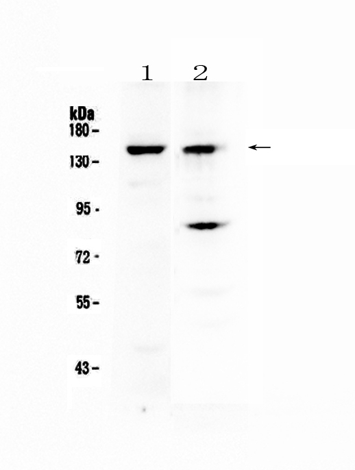
Figure 1. Western blot analysis of FBXL11 using anti-FBXL11 antibody (A03027-1). Electrophoresis was performed on a 5-20% SDS-PAGE gel at 70V (Stacking gel) / 90V (Resolving gel) for 2-3 hours. The sample well of each lane was loaded with 50ug of sample under reducing conditions. Lane 1: mouse liver tissue lysates, Lane 2: human A431 cell lysates. After Electrophoresis, proteins were transferred to a Nitrocellulose membrane at 150mA for 50-90 minutes. Blocked the membrane with 5% Non-fat Milk/ TBS for 1.5 hour at RT. The membrane was incubated with rabbit anti-FBXL11 antigen affinity purified polyclonal antibody (Catalog # A03027-1) at 0.5 microg/mL overnight at 4°C, then washed with TBS-0.1%Tween 3 times with 5 minutes each and probed with a goat anti-rabbit IgG-HRP secondary antibody at a dilution of 1:10000 for 1.5 hour at RT. The signal is developed using an Enhanced Chemiluminescent detection (ECL) kit (Catalog # EK1002) with Tanon 5200 system. A specific band was detected for FBXL11 at approximately 150KD. The expected band size for FBXL11 is at 133KD.
Figure 1. Western blot analysis of FBXL11 using anti-FBXL11 antibody (A03027-1). Electrophoresis was performed on a 5-20% SDS-PAGE gel at 70V (Stacking gel) / 90V (Resolving gel) for 2-3 hours. The sample well of each lane was loaded with 50ug of sample under reducing conditions. Lane 1: mouse liver tissue lysates, Lane 2: human A431 cell lysates. After Electrophoresis, proteins were transferred to a Nitrocellulose membrane at 150mA for 50-90 minutes. Blocked the membrane with 5% Non-fat Milk/ TBS for 1.5 hour at RT. The membrane was incubated with rabbit anti-FBXL11 antigen affinity purified polyclonal antibody (Catalog # A03027-1) at 0.5 microg/mL overnight at 4°C, then washed with TBS-0.1%Tween 3 times with 5 minutes each and probed with a goat anti-rabbit IgG-HRP secondary antibody at a dilution of 1:10000 for 1.5 hour at RT. The signal is developed using an Enhanced Chemiluminescent detection (ECL) kit (Catalog # EK1002) with Tanon 5200 system. A specific band was detected for FBXL11 at approximately 150KD. The expected band size for FBXL11 is at 133KD.
Anti-FBXL11/KDM2A Antibody Picoband(r)
A03027-1-CARRIER-FREE
ApplicationsWestern Blot
Product group Antibodies
ReactivityHuman, Mouse, Rat
TargetKDM2A
Overview
- SupplierBoster Bio
- Product NameAnti-FBXL11/KDM2A Antibody Picoband(r)
- Delivery Days Customer9
- ApplicationsWestern Blot
- CertificationResearch Use Only
- ClonalityPolyclonal
- Concentration500 ug/ml
- Gene ID22992
- Target nameKDM2A
- Target descriptionlysine demethylase 2A
- Target synonymsCXXC8, FBL11, FBL7, FBXL11, JHDM1A, LILINA, lysine-specific demethylase 2A, CXXC-type zinc finger protein 8, F-box and leucine-rich repeat protein 11, F-box/LRR-repeat protein 11, [Histone-H3]-lysine-36 demethylase 1A, jmjC domain-containing histone demethylation protein 1A, jumonji C domain-containing histone demethylase 1A, lysine (K)-specific demethylase 2A
- HostRabbit
- IsotypeIgG
- Protein IDQ9Y2K7
- Protein NameLysine-specific demethylase 2A
- Scientific DescriptionBoster Bio Anti-FBXL11/KDM2A Antibody Picoband® catalog # A03027-1. Tested in WB applications. This antibody reacts with Human, Mouse, Rat. The brand Picoband indicates this is a premium antibody that guarantees superior quality, high affinity, and strong signals with minimal background in Western blot applications. Only our best-performing antibodies are designated as Picoband, ensuring unmatched performance.
- ReactivityHuman, Mouse, Rat
- Storage Instruction-20°C,2°C to 8°C
- UNSPSC12352203
Related products
Product group Antibodies

Anti-KDM2A [RAB-C323]AB01794-1.1-BT
ApplicationsFlow Cytometry, ImmunoFluorescence, ImmunoPrecipitation
ReactivityHuman
TargetKDM2A
- SizePrice
Product group Antibodies

JHDM1A / KDM2A AntibodyLS-C830066
ApplicationsELISA, ImmunoHistoChemistry
ReactivityHuman, Mouse
TargetKDM2A
- SizePrice
Product group Antibodies

Goat anti-KDM2A (aa445-459)EB10899
ApplicationsWestern Blot, ELISA, ImmunoHistoChemistry
ReactivityBovine, Canine, Human, Mouse, Porcine, Rat
TargetKDM2A
- SizePrice
Product group Antibodies
FBXL11 antibody, InternalGTX82550
ApplicationsWestern Blot, ImmunoHistoChemistry, ImmunoHistoChemistry Paraffin
ReactivityHuman
TargetKDM2A
- SizePrice
Product group Antibodies
Anti-JHDM1a/FBXL11 (Center) Antibody102-20485
ApplicationsWestern Blot, ImmunoHistoChemistry, ImmunoHistoChemistry Paraffin
TargetKDM2A
- SizePrice
Product group Antibodies
FBXL11 Polyclonal AntibodyBS-6943R
ApplicationsImmunoFluorescence, ELISA, ImmunoCytoChemistry, ImmunoHistoChemistry, ImmunoHistoChemistry Frozen, ImmunoHistoChemistry Paraffin
ReactivityBovine, Equine, Human, Mouse, Porcine, Rabbit, Rat, Sheep
TargetKDM2A
- SizePrice