


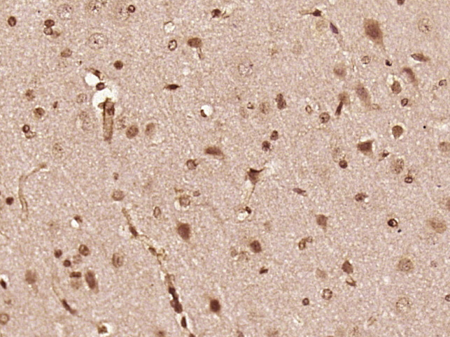
Immunohistochemical staining of human kidney shows strong membranous positivity in cells in tubules.

Immunohistochemical staining of human kidney shows strong membranous positivity in cells in tubules.
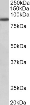
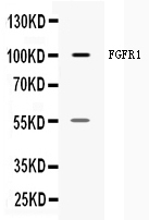
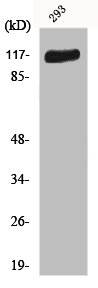
Anti-FGFR1 Antibody
HPA056402
ApplicationsWestern Blot, ImmunoHistoChemistry
Product group Antibodies
ReactivityHuman
TargetFGFR1
Overview
- SupplierAtlas Antibodies
- Product NameAnti-FGFR1 Antibody
- Delivery Days Customer4
- ApplicationsWestern Blot, ImmunoHistoChemistry
- CertificationResearch Use Only
- ClonalityPolyclonal
- ConjugateUnconjugated
- Gene ID2260
- Target nameFGFR1
- Target descriptionfibroblast growth factor receptor 1
- Target synonymsBFGFR, CD331, CEK, ECCL, FGFBR, FGFR-1, FLG, FLT-2, FLT2, HBGFR, HH2, HRTFDS, KAL2, N-SAM, OGD, bFGF-R-1, fibroblast growth factor receptor 1, FGFR1/PLAG1 fusion, FMS-like tyrosine kinase 2, basic fibroblast growth factor receptor 1, fms-related tyrosine kinase 2, heparin-binding growth factor receptor, hydroxyaryl-protein kinase, proto-oncogene c-Fgr
- HostRabbit
- IsotypeIgG
- Protein IDP11362
- Protein NameFibroblast growth factor receptor 1
- Scientific DescriptionRecombinant Protein Epitope Signature Tag (PrEST) antigen sequence
- ReactivityHuman
- Storage Instruction-20°C,2°C to 8°C
- UNSPSC41116161
Datasheet
MSDS
Related products
Product group Antibodies
Anti-FGFR1 AntibodyA82547
ApplicationsWestern Blot, ELISA
ReactivityHuman
- SizePrice
Product group Antibodies

FGFR1 (Phospho-Tyr766) AntibodyABX012438
ApplicationsWestern Blot, ELISA, ImmunoHistoChemistry
- SizePrice
Product group Antibodies

Anti-FGRI [M19B2]Ab02531-1.1
ApplicationsFlow Cytometry, ImmunoFluorescence, ImmunoPrecipitation, Western Blot, ImmunoHistoChemistry, RadioImmunoAssay
ReactivityHuman, Rat
TargetFGFR1
- SizePrice
Product group Antibodies
Anti-Phospho-FGFR1-Y653 Antibody144-50631
ApplicationsWestern Blot
ReactivityHuman, Mouse, Rat
TargetFGFR1
- SizePrice
Product group Antibodies
Anti-FGFR1 Antibody Picoband(r)A00098-CARRIER-FREE
ApplicationsWestern Blot
ReactivityHuman
TargetFGFR1
- SizePrice
Product group Antibodies
ApplicationsImmunoFluorescence, Western Blot, ELISA, ImmunoCytoChemistry, ImmunoHistoChemistry, ImmunoHistoChemistry Frozen, ImmunoHistoChemistry Paraffin
ReactivityBovine, Chicken, Equine, Human, Mouse, Porcine, Rabbit, Rat
TargetFGFR1
- SizePrice
Product group Antibodies
FGFR1 AntibodyCSB-PA002537
ApplicationsImmunoFluorescence, Western Blot, ELISA
ReactivityHuman, Mouse, Rat
TargetFGFR1
- SizePrice