


Immunofluorescent staining of human cell line RT4 shows localization to plasma membrane.
![Lane 1: Marker [kDa] 250, 130, 95, 72, 55, 36, 28, 17, 10 | Lane 2: RT4 | Lane 3: U-251 MG](https://atlasantibodies.s3.amazonaws.com/images/wb/hpa071038-wb-1.jpg "Lane 1: Marker [kDa] 250, 130, 95, 72, 55, 36, 28, 17, 10 | Lane 2: RT4 | Lane 3: U-251 MG")
Immunofluorescent staining of human cell line RT4 shows localization to plasma membrane.
Anti-FLOT2 Antibody
HPA071038
ApplicationsWestern Blot, ImmunoCytoChemistry
Product group Antibodies
ReactivityHuman
TargetFLOT2
Overview
- SupplierAtlas Antibodies
- Product NameAnti-FLOT2 Antibody
- Delivery Days Customer4
- ApplicationsWestern Blot, ImmunoCytoChemistry
- CertificationResearch Use Only
- ClonalityPolyclonal
- ConjugateUnconjugated
- Gene ID2319
- Target nameFLOT2
- Target descriptionflotillin 2
- Target synonymsECS-1, ECS1, ESA, ESA1, M17S1, flotillin-2, Flotillin 2 (epidermal surface antigen 1), epidermal surface antigen, membrane component chromosome 17 surface marker 1, membrane component, chromosome 17, surface marker 1 (35kD protein identified by monoclonal antibody ECS-1)
- HostRabbit
- IsotypeIgG
- Protein IDQ14254
- Protein NameFlotillin-2
- Scientific DescriptionRecombinant Protein Epitope Signature Tag (PrEST) antigen sequence
- ReactivityHuman
- Storage Instruction-20°C,2°C to 8°C
- UNSPSC41116161
Datasheet
MSDS
Related products
Product group Antibodies
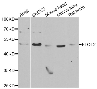
Anti-FLOT2 AntibodyA31440
ApplicationsWestern Blot, ImmunoHistoChemistry
ReactivityHuman, Mouse, Rat
- SizePrice
Product group Antibodies
Anti-FLOT2 (C-term) Antibody102-25148
ApplicationsWestern Blot
TargetFLOT2
- SizePrice
Product group Antibodies

FLOT2 / Flotillin 2 AntibodyLS-C830604
ApplicationsWestern Blot, ELISA, ImmunoHistoChemistry
ReactivityHuman, Mouse, Rat
TargetFLOT2
- SizePrice
Product group Antibodies
Flotillin 2 Recombinant AntibodyBSM-61102R
ApplicationsImmunoPrecipitation, Western Blot
ReactivityHuman, Mouse, Rat
TargetFLOT2
- SizePrice
Product group Antibodies
FLOT2 AntibodyCSB-PA002545
ApplicationsWestern Blot, ELISA
ReactivityHuman, Mouse, Rat
TargetFLOT2
- SizePrice
Product group Antibodies

ApplicationsWestern Blot, ELISA, ImmunoHistoChemistry
ReactivityHuman
TargetFLOT2
- SizePrice
Product group Antibodies
ApplicationsImmunoPrecipitation, Western Blot, ImmunoCytoChemistry, ImmunoHistoChemistry
TargetFLOT2
- SizePrice
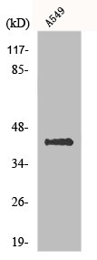
![293T whole cell extracts and 293T exosome extract (2.6 μg) were separated by 10% SDS-PAGE, and the membrane was blotted with Flotillin 2 antibody [C3], C-term (GTX100279) diluted at 1:250. The HRP-conjugated anti-rabbit IgG antibody (GTX213110-01) was used to detect the primary antibody, and the signal was developed with Trident ECL plus-Enhanced.](https://www.genetex.com/upload/website/prouct_img/normal/GTX100279/GTX100279_39443_20190607_WB_Fraction_w_23060100_908.webp)
Product group Antibodies
Flotillin 2 antibody [C3], C-termGTX100279
ApplicationsImmunoFluorescence, ImmunoPrecipitation, Western Blot, ImmunoCytoChemistry, ImmunoHistoChemistry, ImmunoHistoChemistry Paraffin
ReactivityHuman, Mouse, Rat
TargetFLOT2
- SizePrice