


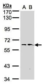
Figure 1. Western blot analysis of FMO1 using anti-FMO1 antibody (PB9589). Electrophoresis was performed on a 5-20% SDS-PAGE gel at 70V (Stacking gel) / 90V (Resolving gel) for 2-3 hours. Lane 1: Rat Liver Tissue Lysate at 50ug, Lane 2: Mouse Liver Tissue Lysate at 50ug, Lane 3: Rat Kidney Tissue Lysate at 50ug, Lane 4: Mouse Kidney Tissue Lysate at 50ug, Lane 5: SMMC Whole Cell Lysate at 40ug. After electrophoresis, proteins were transferred to a nitrocellulose membrane at 150 mA for 50-90 minutes. Blocked the membrane with 5% non-fat milk/TBS for 1.5 hour at RT. The membrane was incubated with rabbit anti-FMO1 antigen affinity purified polyclonal antibody (Catalog # PB9589) at 0.5 microg/mL overnight at 4°C, then washed with TBS-0.1%Tween 3 times with 5 minutes each and probed with a goat anti-rabbit IgG-HRP secondary antibody at a dilution of 1:5000 for 1.5 hour at RT. The signal is developed using an Enhanced Chemiluminescent detection (ECL) kit (Catalog # EK1002) with Tanon 5200 system. A specific band was detected for FMO1 at approximately 60 kDa. The expected band size for FMO1 is at 60 kDa.
Figure 1. Western blot analysis of FMO1 using anti-FMO1 antibody (PB9589). Electrophoresis was performed on a 5-20% SDS-PAGE gel at 70V (Stacking gel) / 90V (Resolving gel) for 2-3 hours. Lane 1: Rat Liver Tissue Lysate at 50ug, Lane 2: Mouse Liver Tissue Lysate at 50ug, Lane 3: Rat Kidney Tissue Lysate at 50ug, Lane 4: Mouse Kidney Tissue Lysate at 50ug, Lane 5: SMMC Whole Cell Lysate at 40ug. After electrophoresis, proteins were transferred to a nitrocellulose membrane at 150 mA for 50-90 minutes. Blocked the membrane with 5% non-fat milk/TBS for 1.5 hour at RT. The membrane was incubated with rabbit anti-FMO1 antigen affinity purified polyclonal antibody (Catalog # PB9589) at 0.5 microg/mL overnight at 4°C, then washed with TBS-0.1%Tween 3 times with 5 minutes each and probed with a goat anti-rabbit IgG-HRP secondary antibody at a dilution of 1:5000 for 1.5 hour at RT. The signal is developed using an Enhanced Chemiluminescent detection (ECL) kit (Catalog # EK1002) with Tanon 5200 system. A specific band was detected for FMO1 at approximately 60 kDa. The expected band size for FMO1 is at 60 kDa.
Anti-FMO1 Antibody Picoband(r)
PB9589-CARRIER-FREE
ApplicationsWestern Blot
Product group Antibodies
ReactivityHamster, Human, Mouse, Rat
TargetFMO1
Overview
- SupplierBoster Bio
- Product NameAnti-FMO1 Antibody Picoband(r)
- Delivery Days Customer9
- Application Supplier NoteTested Species: In-house tested species with positive results. Other applications have not been tested. Optimal dilutions should be determined by end users.
- ApplicationsWestern Blot
- CertificationResearch Use Only
- ClonalityPolyclonal
- Concentration500 ug/ml
- Gene ID2326
- Target nameFMO1
- Target descriptionflavin containing dimethylaniline monoxygenase 1
- Target synonymsflavin-containing monooxygenase 1, FMO 1, Flavin-containing monooxygenase 1 (fetal liver), dimethylaniline monooxygenase [N-oxide-forming] 1, dimethylaniline oxidase 1, fetal hepatic flavin-containing monooxygenase 1, flavin containing monooxygenase 1, trimethylamine monooxygenase
- HostRabbit
- IsotypeIgG
- Protein IDQ01740
- Protein NameFlavin-containing monooxygenase 1
- Scientific DescriptionBoster Bio Anti-FMO1 Antibody Picoband® catalog # PB9589. Tested in WB applications. This antibody reacts with Human, Mouse, Rat. The brand Picoband indicates this is a premium antibody that guarantees superior quality, high affinity, and strong signals with minimal background in Western blot applications. Only our best-performing antibodies are designated as Picoband, ensuring unmatched performance.
- ReactivityHamster, Human, Mouse, Rat
- Storage Instruction-20°C,2°C to 8°C
- UNSPSC12352203
Related products
Product group Antibodies
FMO1 AntibodyCSB-PA008746ESR2HU
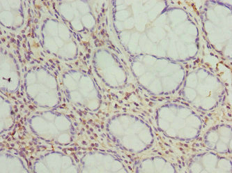
ApplicationsELISA, ImmunoHistoChemistry
ReactivityHuman
TargetFMO1
- SizePrice

Product group Antibodies
Anti-FMO1 AntibodyHPA023680
ApplicationsImmunoHistoChemistry
ReactivityHuman
TargetFMO1
- SizePrice
Product group Antibodies

FMO1 AntibodyLS-C675767
ApplicationsELISA, ImmunoHistoChemistry, ImmunoHistoChemistry Paraffin
ReactivityHuman
TargetFMO1
- SizePrice
Product group Antibodies
ApplicationsImmunoPrecipitation, Western Blot, ImmunoCytoChemistry, ImmunoHistoChemistry
ReactivityPorcine
TargetFMO1
- SizePrice
Product group Antibodies
FMO1 AntibodyPACO44477
ApplicationsELISA, ImmunoHistoChemistry
ReactivityHuman
TargetFMO1
- SizePrice
Product group Antibodies
FMO1 antibody [N1C2]GTX104172
ApplicationsWestern Blot
ReactivityHuman
TargetFMO1
- SizePrice
Product group Antibodies
Anti-FMO1 Antibody107-10040
ApplicationsWestern Blot
ReactivityHuman
TargetFMO1
- SizePrice