


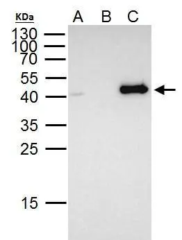
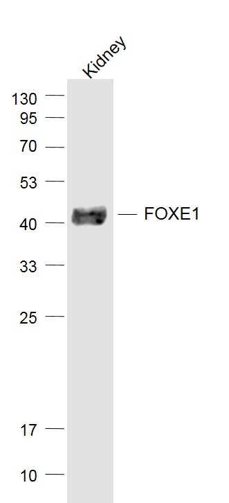
Figure 1. Western blot analysis of FOXE1 using anti-FOXE1 antibody (A02831-5). Electrophoresis was performed on a 5-20% SDS-PAGE gel at 70V (Stacking gel) / 90V (Resolving gel) for 2-3 hours. The sample well of each lane was loaded with 50ug of sample under reducing conditions. Lane 1: human Hela whole cell lysates. After Electrophoresis, proteins were transferred to a Nitrocellulose membrane at 150mA for 50-90 minutes. Blocked the membrane with 5% Non-fat Milk/ TBS for 1.5 hour at RT. The membrane was incubated with rabbit anti-FOXE1 antigen affinity purified polyclonal antibody (Catalog # A02831-5) at 0.5 microg/mL overnight at 4°C, then washed with TBS-0.1%Tween 3 times with 5 minutes each and probed with a goat anti-rabbit IgG-HRP secondary antibody at a dilution of 1:5000 for 1.5 hour at RT. The signal is developed using an Enhanced Chemiluminescent detection (ECL) kit (Catalog # EK1002) with Tanon 5200 system. A specific band was detected for FOXE1 at approximately 41KD. The expected band size for FOXE1 is at 38KD.
. Overlay histogram showing SiHa cells stained with A02831-5 (Blue line). To facilitate intracellular staining, cells were fixed with 4% paraformaldehyde and permeabilized with permeabilization buffer. The cells were blocked with 10% normal goat serum. And then incubated with rabbit anti-FOXE1 Antibody (A02831-5, 1microg/1x106 cells) for 30 min at 20°C. DyLight®488 conjugated goat anti-rabbit IgG (BA1127, 5-10microg/1x106 cells) was used as secondary antibody for 30 minutes at 20°C. Isotype control antibody (Green line) was rabbit IgG (1microg/1x106) used under the same conditions. Unlabelled sample without incubation with primary antibody and secondary antibody (Red line) was used as a blank control.")
Figure 1. Western blot analysis of FOXE1 using anti-FOXE1 antibody (A02831-5). Electrophoresis was performed on a 5-20% SDS-PAGE gel at 70V (Stacking gel) / 90V (Resolving gel) for 2-3 hours. The sample well of each lane was loaded with 50ug of sample under reducing conditions. Lane 1: human Hela whole cell lysates. After Electrophoresis, proteins were transferred to a Nitrocellulose membrane at 150mA for 50-90 minutes. Blocked the membrane with 5% Non-fat Milk/ TBS for 1.5 hour at RT. The membrane was incubated with rabbit anti-FOXE1 antigen affinity purified polyclonal antibody (Catalog # A02831-5) at 0.5 microg/mL overnight at 4°C, then washed with TBS-0.1%Tween 3 times with 5 minutes each and probed with a goat anti-rabbit IgG-HRP secondary antibody at a dilution of 1:5000 for 1.5 hour at RT. The signal is developed using an Enhanced Chemiluminescent detection (ECL) kit (Catalog # EK1002) with Tanon 5200 system. A specific band was detected for FOXE1 at approximately 41KD. The expected band size for FOXE1 is at 38KD.
Anti-FOXE1 Picoband(r) Antibody
A02831-5-CARRIER-FREE
ApplicationsFlow Cytometry, Western Blot, ELISA
Product group Antibodies
ReactivityHuman
TargetFOXE1
Overview
- SupplierBoster Bio
- Product NameAnti-FOXE1 Picoband(r) Antibody
- Delivery Days Customer9
- ApplicationsFlow Cytometry, Western Blot, ELISA
- CertificationResearch Use Only
- ClonalityPolyclonal
- Concentration500 ug/ml
- Gene ID2304
- Target nameFOXE1
- Target descriptionforkhead box E1
- Target synonymsBAMLAZ, FKHL15, FOXE2, HFKH4, HFKL5, NMTC4, TITF2, TTF-2, TTF2, forkhead box protein E1, HNF-3/fork head-like protein 5, forkhead box protein E2, forkhead, drosophila, homolog-like 15, forkhead-related protein FKHL15, thyroid transcription factor 2
- HostRabbit
- IsotypeIgG
- Protein IDO00358
- Protein NameForkhead box protein E1
- Scientific DescriptionBoster Bio Anti-FOXE1 Picoband® Antibody catalog # A02831-5. Tested in ELISA, Flow Cytometry, WB applications. This antibody reacts with Human. The brand Picoband indicates this is a premium antibody that guarantees superior quality, high affinity, and strong signals with minimal background in Western blot applications. Only our best-performing antibodies are designated as Picoband, ensuring unmatched performance.
- ReactivityHuman
- Storage Instruction-20°C,2°C to 8°C
- UNSPSC12352203
Related products
Product group Antibodies
FOXE1 AntibodyCSB-PA008314
ApplicationsImmunoFluorescence, ELISA
ReactivityHuman, Mouse, Rat
TargetFOXE1
- SizePrice
Product group Antibodies
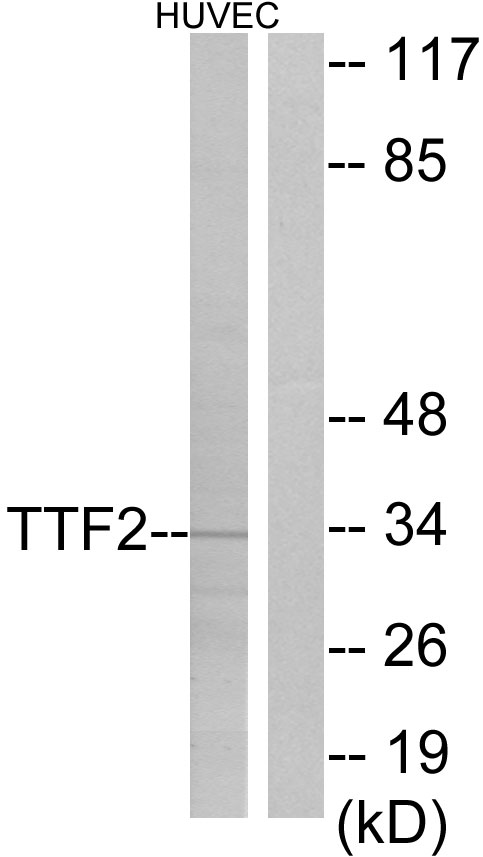
Anti-TTF2 AntibodyA97875
ApplicationsWestern Blot, ELISA
ReactivityHuman, Mouse, Rat
- SizePrice
Product group Antibodies

FOXE2 / FOXE1 AntibodyLS-C747721
ApplicationsWestern Blot
ReactivityHuman
TargetFOXE1
- SizePrice
Product group Antibodies

Goat anti-FOXE1 / TTF2EB06011
ApplicationsELISA, ImmunoHistoChemistry
ReactivityHuman
TargetFOXE1
- SizePrice
Product group Antibodies
Foxe1 Polyclonal AntibodyCAC08156
ApplicationsImmunoFluorescence, Western Blot, ELISA
ReactivityMouse
TargetFOXE1
- SizePrice
Product group Antibodies
FOXE1 antibodyGTX100275
ApplicationsImmunoPrecipitation, Western Blot
ReactivityHuman
TargetFOXE1
- SizePrice
Product group Antibodies
References
FOXE1 Polyclonal AntibodyBS-0446R
ApplicationsImmunoFluorescence, Western Blot, ELISA, ImmunoCytoChemistry, ImmunoHistoChemistry, ImmunoHistoChemistry Frozen, ImmunoHistoChemistry Paraffin
ReactivityBovine, Canine, Chicken, Human, Mouse, Porcine, Rat
TargetFOXE1
- SizePrice